


ABSTRACT
Background and Aim: Houseflies (Musca domestica) are recognized mechanical vectors of foodborne pathogens and antimicrobial-resistant bacteria, particularly in environments with intensive food-handling. Their role in disseminating virulent and multidrug-resistant (MDR) Escherichia coli at the food–environment–human interface remains underexplored in Northern Thailand. This study aimed to determine the prevalence, phylogenetic distribution, virulence gene carriage, antimicrobial resistance profiles, and resistance determinants of E. coli isolated from houseflies collected at food markets in Phayao Province.
Materials and Methods: A cross-sectional surveillance study was conducted between June and November 2023 in Muang District, Phayao Province. A total of 350 houseflies were collected from meat, vegetable, and cooked-food markets using sticky traps. E. coli isolates were identified by culture and polymerase chain reaction (PCR) targeting the uidA gene. Phylogenetic grouping was performed using chuA, yjaA, and TSPE4.C2. Virulence genes associated with diarrheagenic E. coli were detected by PCR. Antimicrobial susceptibility testing was conducted using the disc diffusion method against 12 antibiotics representing eight antimicrobial classes. Multidrug resistance was defined as resistance to three or more antimicrobial classes. Resistance-associated genes were screened by PCR. Associations were analyzed using the chi-square test.
Results: Overall, 106 E. coli isolates (30.3%) were recovered, with the highest prevalence in meat markets (39.5%) (p < 0.0001). Phylogroup A predominated (49.1%), followed by D (19.8%), B1 (18.9%), and B2 (12.3%). Virulence genes were detected in 69.8% of isolates, with bfpA being the most prevalent (26.4%). Universal resistance to penicillin G and erythromycin was observed, while high resistance rates were noted for ampicillin (66.0%) and tetracycline (35.8%). MDR was identified in 46.2% of isolates and was significantly more common in meat markets (p = 0.0225). The highest MDR prevalence occurred in phylogroup D (57.1%). The most frequently detected resistance genes were blaSHV, ereA, and tetA.
Conclusion: Houseflies from food markets in Northern Thailand harbor virulent and MDR E. coli, highlighting their role as environmental sentinels and potential disseminators of antimicrobial resistance. These findings underscore the need for improved market hygiene and incorporation of insect vectors into One Health–based antimicrobial resistance surveillance strategies.
Keywords: antimicrobial resistance, Escherichia coli, food markets, houseflies, multidrug resistance, Northern Thailand, One Health, virulence genes.
INTRODUCTION
Antimicrobial resistance (AMR) has emerged as a critical global public health challenge and has received considerable attention from the World Health Organization (WHO) [1]. The increasing prevalence of pathogens exhibiting multidrug-resistant (MDR) phenotypes represents a major threat, as it substantially complicates the management of common infections and contributes to elevated morbidity, mortality, and healthcare costs [2, 3]. Owing to its far-reaching consequences for public health and global economic stability, the WHO has identified AMR as a strategic priority within the framework of the Sustainable Development Goals [4, 5]. Effective mitigation of AMR requires integrated approaches, including the establishment of robust surveillance systems, the promotion of targeted research, and the implementation of evidence-based public health interventions. Escherichia coli is a Gram-negative bacillus that forms part of the normal gastrointestinal microbiota of humans and other warm-blooded animals; however, pathogenic strains are capable of causing gastrointestinal disease as well as extraintestinal infections, such as urinary tract infections and septicemia [6]. The rising prevalence of MDR E. coli is therefore a major public health concern, as it restricts therapeutic options and increases the likelihood of treatment failure [7].
Houseflies are recognized mechanical vectors of a wide range of pathogens, including viruses, fungi, and bacteria such as E. coli. These insects can acquire and disseminate microorganisms throughout their life cycle, particularly in environments with inadequate sanitation [8]. Their feeding and breeding activities are closely linked to food markets, animal production systems, and waste disposal sites, where they can mechanically transfer pathogenic and AMR bacteria from contaminated substrates to food and food-contact surfaces [9]. Poor hygiene conditions facilitate housefly proliferation, thereby increasing the risk of pathogen transmission to humans [10]. The role of houseflies in the dissemination of AMR bacteria underscores their epidemiological importance and highlights the need for surveillance of MDR E. coli in food market environments, where the risk of human exposure is particularly high.
Despite the growing recognition of AMR as a major global health threat, surveillance efforts have largely focused on clinical, livestock, and food samples, with comparatively limited attention given to environmental vectors operating at the human–food interface. In particular, the role of houseflies as mechanical carriers of MDR E. coli in fresh-food market environments remains insufficiently characterized, especially in Northern Thailand. Existing studies from Thailand and neighboring regions have primarily reported prevalence and phenotypic antimicrobial susceptibility, with limited integration of phylogenetic background, virulence determinants, and resistance gene profiles within the same analytical framework. Moreover, comparative data across different market types (meat, vegetable, and cooked-food markets) are scarce, constraining risk assessment of differential exposure scenarios. The absence of region-specific baseline data that simultaneously link MDR phenotypes, virulence-associated genes, and phylogenetic distribution of E. coli in housefly populations represents a critical knowledge gap for environmental AMR surveillance and One Health–oriented interventions.
This study aimed to determine the prevalence of E. coli isolated from houseflies collected at meat, vegetable, and cooked-food markets in Muang District, Phayao Province, Northern Thailand, and to characterize their phylogenetic distribution, virulence gene profiles, antimicrobial resistance patterns, and MDR status. In addition, this study sought to identify resistance-associated genes and to evaluate the associations between market type, phylogenetic group, virulence carriage, and MDR phenotypes. By generating integrated baseline data from a high-risk food market setting, the study aims to contribute evidence to support AMR surveillance and control strategies within a One Health framework.
MATERIALS AND METHODS
Ethical approval
This study did not require formal ethical approval from an Institutional Animal Ethics Committee or Institutional Review Board, as it exclusively involved the collection of houseflies (Musca domestica), which are invertebrates and not subject to animal welfare regulations under international guidelines (e.g., those from the World Organization for Animal Health or local Thai regulations). No vertebrate animals, human participants, or procedures causing pain or distress were used. The study was conducted in accordance with general principles of humane insect collection and biosafety protocols at the University of Phayao. This study on M. domestica was reviewed and approved by the Institutional Animal Care and Use Committee of the University of Phayao, Thailand (Ethical Approval No. 640104039).
Study period and location
This cross-sectional surveillance study was conducted from June to November 2023 in urban and peri-urban market settings in Muang District, Phayao Province, Northern Thailand.
Sample collection
Housefly samples were collected from local meat, vegetable, and cooked-food markets selected based on public accessibility, active food-handling activities, and their relevance as high-contact environments with potential environmental contamination and human exposure.
Sampling intensity was adjusted according to market size, with five traps deployed per sampling event in cooked-food markets (small), ten in vegetable markets (medium), and fifteen in meat markets (large). In total, 350 houseflies were collected using the sticky trap method, as previously described [11]. Sterile adhesive strips were placed within market areas, and captured houseflies were transferred individually into sterile containers using forceps. Samples were transported to the laboratory and euthanized at −20°C for 1–2 h. Morphological identification was performed using a stereomicroscope to confirm species identity as M. domestica [12]. A single housefly was then placed in a test tube containing 2 mL of alkaline peptone water (APW) medium (HiMedia Laboratories Pvt. Ltd., Mumbai, India, Cat. No. M618-500G), vortexed, and incubated at 37°C for 16–18 h for whole-fly enrichment without separation of external and internal compartments.
Isolation of E. coli
A sterile loopful of enriched APW culture was streaked onto eosin methylene blue agar (HiMedia Laboratories Pvt. Ltd., Cat. No.M317-500G) and incubated at 37°C for 24 h. Presumptive E. coli colonies exhibiting a metallic sheen with a dark center were selected and subjected to biochemical and morphological characterization, including Gram staining, sugar fermentation, methyl red, Voges–Proskauer, indole, and citrate tests, as previously described [13]. Confirmed colonies were cultured and stored on nutrient agar (NA) medium (HiMedia Laboratories Pvt. Ltd., Cat. No. M001-500G) and incubated at 37°C for 24 h for further analyses.
DNA extraction
Genomic DNA was extracted using the boiling method, as previously described [14]. A single colony of E. coli grown on NA medium was suspended in 100 μL of sterile water in a 1.5 mL microcentrifuge tube. Samples were heated at 100°C for 10 min, cooled on ice for 5 min, and centrifuged at 10,000 rpm for 10 min. The supernatant was transferred to a new microcentrifuge tube and stored at −20°C until use.
Molecular confirmation and phylogenetic grouping
All isolates were molecularly confirmed as E. coli by polymerase chain reaction (PCR) targeting the uidA (β-glucuronidase) gene, following Heijnen and Medema [15] with minor modifications. PCR reactions (25 μL) were prepared using OnePCR™ Ultra (Bio-Helix Co., Ltd., Keelung, Taiwan, Cat. No. 1BHC-MBA01-0100), uidA-F/uidA-R primers (Supplementary Table 1), DNA template, and sterile water. PCR products were analyzed by agarose gel electrophoresis and visualized under UV light using a gel documentation system (BIS 303 PC; DNR Bio-Imaging Systems Ltd., Jerusalem, Israel). A 100 bp DNA marker (OmniMARK 100 RTU., Bio-Helix Co., Ltd., Cat. No. DM101-0100) was used for size estimation. Amplicons were sequenced by Macrogen Inc. (Seoul, South Korea) and analyzed using BLAST in the NCBI GenBank database.
Phylogenetic grouping was performed by multiplex PCR targeting the chuA and yjaA genes and the DNA fragment TSPE4.C2, as described by Clermont et al. [16]. PCR conditions and downstream analyses followed the same procedures as described above.
Detection of virulence genes
Virulence genes associated with diarrheagenic E. coli were detected by PCR using primers and conditions described previously [7]. The target genes included aggR, stx1, stx2, astA, estp, esth, elt, bfpA, eae, and invE (Supplementary Table 1).
Antimicrobial susceptibility testing
Antimicrobial susceptibility was evaluated using the disk diffusion method in accordance with Clinical and Laboratory Standards Institute guidelines (CLSI M100-S23, 2018). Twelve antibiotics representing eight antimicrobial classes were tested: aminoglycosides (gentamicin, streptomycin), penicillins/β-lactams (penicillin G, ampicillin), carbapenems (imipenem, meropenem), tetracyclines (tetracycline, tigecycline), phenicols (chloramphenicol), fluoroquinolones (ciprofloxacin), macrolides (erythromycin), and sulfonamides (trimethoprim–sulfamethoxazole) (HiMedia Laboratories Pvt. Ltd., Cat. No. SD170, SD031, SD028, SD002, SD073, SD727, SD037, SD278, SD006, SD060, SD013, and SD010, respectively). Mueller–Hinton agar plates (HiMedia Laboratories Pvt. Ltd., Cat. No. M173) were inoculated with a 0.5 McFarland standardized suspension and incubated at 37°C for 18–24 h. Inhibition zones were measured in millimeters. E. coli TISTR 073 served as the quality control strain. Isolates resistant to one or more antibiotics in three or more antimicrobial classes were classified as MDR [17, 18].
Detection of antimicrobial resistance genes
Phenotypically resistant E. coli isolates were screened for resistance-associated genes by PCR. Target genes included acc(3)-IV, aac(6)-Ib-cr, aadA1, CITM, blaSHV, catA1, ereA, sul1, and tetA. Primer sequences and expected amplicon sizes are provided in Supplementary Table 1. PCR conditions followed previously described protocols [7, 10, 15, 16, 19, 20].
Statistical analysis
Descriptive data are presented as frequencies and percentages. Differences in categorical variables, including E. coli prevalence, virulence gene positivity, MDR phenotypes, and AMR gene distribution among market types, were assessed using the chi-square test. Associations between phylogenetic groups and virulence status, as well as between phylogenetic groups and MDR status, were also evaluated. Statistical analyses were performed using GraphPad Prism version 5.00 for Windows (GraphPad Software Inc., San Diego, CA, USA), and p ≤ 0.05 was considered statistically significant.
RESULTS
Prevalence and phylogroup distribution of E. coli isolates
In this study, 106 (30.3%) E. coli strains were isolated from 350 housefly samples and confirmed by PCR analysis. The highest prevalence was observed in the meat market (39.5%), followed by the cooked-food market (26.3%) and the vegetable market (14.0%) (Table 1). A statistically significant difference in E. coli prevalence was detected among market types (p < 0.0001). Phylogenetic analysis revealed that phylogroup A was predominant (52 isolates; 49.1%), followed by phylogroups D (21 isolates; 19.8%), B1 (20 isolates; 18.9%), and B2 (13 isolates; 12.3%) (Table 1).
| Sampling area | Number of samples collected | E. coli-positive samples, n (%) | A, n (%) | B1, n (%) | B2, n (%) | D, n (%) |
|---|---|---|---|---|---|---|
| Meat market | 200 | 79 (39.5) | 42 (53.2) | 11 (13.9) | 7 (8.9) | 19 (24.1) |
| Vegetable market | 100 | 14 (14.0) | 7 (50.0) | 5 (35.7) | 0 (0.0) | 2 (14.3) |
| Cooked-food market | 50 | 13 (26.3) | 3 (23.1) | 4 (30.8) | 6 (46.2) | 0 (0.0) |
| Total | 350 | 106 (30.3) | 52 (49.1) | 20 (18.9) | 13 (12.3) | 21 (19.8) |
Table 1. Prevalence and occurrence rate of phylogenetic groups of Escherichia coli isolates obtained from houseflies at various markets in Phayao Province, Northern Thailand.
| Sampling area | Number of samples collected | E. coli-positive samples, n (%) | A, n (%) | B1, n (%) | B2, n (%) | D, n (%) |
|---|---|---|---|---|---|---|
| Meat market | 200 | 79 (39.5) | 42 (53.2) | 11 (13.9) | 7 (8.9) | 19 (24.1) |
| Vegetable market | 100 | 14 (14.0) | 7 (50.0) | 5 (35.7) | 0 (0.0) | 2 (14.3) |
| Cooked-food market | 50 | 13 (26.3) | 3 (23.1) | 4 (30.8) | 6 (46.2) | 0 (0.0) |
| Total | 350 | 106 (30.3) | 52 (49.1) | 20 (18.9) | 13 (12.3) | 21 (19.8) |
n = Number of isolates, % = Percentage within E. coli-positive samples, A, B1, B2, and D = Phylogenetic groups of E. coli. p-value: <0.0001.
Distribution of virulence genes in E. coli isolates
All E. coli isolates were screened for 10 virulence genes associated with diarrheagenic pathotypes. Overall, 74 isolates (69.8%) were positive for at least one virulence gene (Table 2). The most prevalent gene was bfpA (26.4%; 28/106), followed by astA (12.3%; 13/106), stx1 (7.5%; 8/106), esth (4.7%; 5/106), eae (2.8%; 3/106), and estp (0.9%; 1/106). In contrast, elt, invE, and stx2 were not detected. No significant difference was observed in the distribution of virulence gene-positive isolates among market types (p = 0.057). Isolates carrying two virulence genes accounted for 15.1% (16/106), with four observed combinations, the most common being aggR–astA (9.4%; 10/106), followed by aggR–bfpA (3.8%; 4/106), astA–esth (0.9%; 1/106), and bfpA–stx (0.9%; 1/106).
| Virulence profiles | Meat market (n = 79) | Vegetable market (n = 14) | Cooked-food market (n = 13) | Total (n = 106) | p-value |
|---|---|---|---|---|---|
| Negative | 24 (30.4) | 7 (50.0) | 1 (7.7) | 32 (30.2) | 0.0570 |
| Positive virulence | 55 (69.6) | 7 (50.0) | 12 (92.3) | 74 (69.8) | |
| One gene | |||||
| astA | 13 (16.5) | 0 (0.0) | 0 (0.0) | 13 (12.3) | |
| bfpA | 24 (30.4) | 1 (7.1) | 3 (23.1) | 28 (26.4) | |
| eae | 3 (3.8) | 0 (0.0) | 0 (0.0) | 3 (2.8) | |
| esth | 5 (6.3) | 0 (0.0) | 0 (0.0) | 5 (4.7) | |
| estp | 0 (0.0) | 0 (0.0) | 1 (7.7) | 1 (0.9) | |
| stx1 | 4 (5.1) | 4 (28.6) | 0 (0.0) | 8 (7.5) | |
| Two genes | |||||
| aggR–astA | 5 (6.3) | 1 (7.1) | 4 (30.8) | 10 (9.4) | |
| aggR–bfpA | 0 (0.0) | 0 (0.0) | 4 (30.8) | 4 (3.8) | |
| astA–esth | 1 (1.3) | 0 (0.0) | 0 (0.0) | 1 (0.9) | |
| bfpA–stx | 0 (0.0) | 1 (7.1) | 0 (0.0) | 1 (0.9) |
Table 2. Virulence gene profiles of Escherichia coli isolated from houseflies in various markets in Phayao Province, Northern Thailand.
| Virulence profiles | Meat market (n = 79) | Vegetable market (n = 14) | Cooked-food market (n = 13) | Total (n = 106) | p-value |
|---|---|---|---|---|---|
| Negative | 24 (30.4) | 7 (50.0) | 1 (7.7) | 32 (30.2) | 0.0570 |
| Positive virulence | 55 (69.6) | 7 (50.0) | 12 (92.3) | 74 (69.8) | |
| One gene | |||||
| astA | 13 (16.5) | 0 (0.0) | 0 (0.0) | 13 (12.3) | |
| bfpA | 24 (30.4) | 1 (7.1) | 3 (23.1) | 28 (26.4) | |
| eae | 3 (3.8) | 0 (0.0) | 0 (0.0) | 3 (2.8) | |
| esth | 5 (6.3) | 0 (0.0) | 0 (0.0) | 5 (4.7) | |
| estp | 0 (0.0) | 0 (0.0) | 1 (7.7) | 1 (0.9) | |
| stx1 | 4 (5.1) | 4 (28.6) | 0 (0.0) | 8 (7.5) | |
| Two genes | |||||
| aggR–astA | 5 (6.3) | 1 (7.1) | 4 (30.8) | 10 (9.4) | |
| aggR–bfpA | 0 (0.0) | 0 (0.0) | 4 (30.8) | 4 (3.8) | |
| astA–esth | 1 (1.3) | 0 (0.0) | 0 (0.0) | 1 (0.9) | |
| bfpA–stx | 0 (0.0) | 1 (7.1) | 0 (0.0) | 1 (0.9) |
n = Number of Escherichia coli isolates, % = Percentage within each market group, astA = enteroaggregative heat-stable toxin gene, bfpA = bundle-forming pilus gene, eae = intimin gene, esth = heat-stable enterotoxin gene (human type), estp = heat-stable enterotoxin gene (porcine type), stx1 = Shiga toxin 1 gene, aggR = transcriptional activator gene. p-values were calculated using the chi-square test for comparisons of the distribution of virulence gene-positive E. coli isolates among market types.
Phylogenetic distribution of virulence-associated gene profiles
Virulence gene profiles varied across phylogenetic groups (Supplementary Table 2). Phylogroup A exhibited the greatest diversity of virulence-associated genes, with bfpA being the most prevalent profile (36.4%; 12/33), whereas rare profiles such as bfpA–stx were least frequent (3.0%; 1/33). In phylogroup B1, bfpA predominated (35.7%; 5/14), while stx1 occurred at the lowest frequency (7.1%; 1/14). Phylogroup B2 showed marked dominance of bfpA (63.6%; 7/11), representing the highest single-gene prevalence among all groups, whereas estp was detected infrequently and exclusively in this group (9.1%; 1/11). Phylogroup D displayed a more even distribution of virulence profiles, with bfpA, astA, stx1, and aggR–astA representing the most common patterns (18.8%–25.0%; 3–4/16), while eae was the least prevalent (6.3%; 1/16). No significant association was found between phylogenetic group and overall virulence gene positivity (p = 0.4314).
Antibiotic resistance phenotype of E. coli isolates
Phenotypic antimicrobial resistance was widely observed among E. coli isolates from all markets. All 106 isolates were tested against 12 antibiotics representing eight antimicrobial classes. Complete resistance to penicillin G (100%) and erythromycin (100%) was observed in all isolates (Table 3). High resistance rates were recorded for ampicillin (66.0%; 70/106), tetracycline (35.8%; 38/106), and chloramphenicol (17.0%; 18/106). Lower resistance rates were noted for gentamicin (11.3%; 12/106), streptomycin (11.3%; 12/106), sulfonamide (11.3%; 12/106), and ciprofloxacin (2.8%; 3/106). No resistance was detected to imipenem, meropenem, or tigecycline.
| Drug classes | Antibiotics | Meat (n = 79) | Vegetable market (n = 14) | Cooked-food market (n = 13) | Total (n = 106) |
|---|---|---|---|---|---|
| Aminoglycosides | Gentamicin | 7 (8.9) | 0 (0.0) | 5 (38.5) | 12 (11.3) |
| Streptomycin | 12 (15.2) | 0 (0.0) | 0 (0.0) | 12 (11.3) | |
| Penicillin | Penicillin G | 79 (100.0) | 14 (100.0) | 13 (100.0) | 106 (100.0) |
| Ampicillin | 55 (69.9) | 7 (50.0) | 8 (61.5) | 70 (66.0) | |
| Carbapenem | Imipenem | 0 (0.0) | 0 (0.0) | 0 (0.0) | 0 (0.0) |
| Meropenem | 0 (0.0) | 0 (0.0) | 0 (0.0) | 0 (0.0) | |
| Tetracycline | Tetracycline | 33 (41.8) | 0 (0.0) | 5 (38.5) | 38 (35.8) |
| Tigecycline | 0 (0.0) | 0 (0.0) | 0 (0.0) | 0 (0.0) | |
| Phenicol | Chloramphenicol | 13 (16.5) | 0 (0.0) | 5 (38.5) | 18 (17.0) |
| Fluoroquinolone | Ciprofloxacin | 3 (3.8) | 0 (0.0) | 0 (0.0) | 3 (2.8) |
| Macrolide | Erythromycin | 79 (100.0) | 14 (100.0) | 13 (100.0) | 106 (100.0) |
| Sulfonamides | Trimethoprim/sulfamethoxazole | 10 (12.7) | 2 (14.3) | 0 (0.0) | 12 (11.3) |
Table 3. Antibiotic resistance profile of Escherichia coli isolates.
| Drug classes | Antibiotics | Meat (n = 79) | Vegetable market (n = 14) | Cooked-food market (n = 13) | Total (n = 106) |
|---|---|---|---|---|---|
| Aminoglycosides | Gentamicin | 7 (8.9) | 0 (0.0) | 5 (38.5) | 12 (11.3) |
| Streptomycin | 12 (15.2) | 0 (0.0) | 0 (0.0) | 12 (11.3) | |
| Penicillin | Penicillin G | 79 (100.0) | 14 (100.0) | 13 (100.0) | 106 (100.0) |
| Ampicillin | 55 (69.9) | 7 (50.0) | 8 (61.5) | 70 (66.0) | |
| Carbapenem | Imipenem | 0 (0.0) | 0 (0.0) | 0 (0.0) | 0 (0.0) |
| Meropenem | 0 (0.0) | 0 (0.0) | 0 (0.0) | 0 (0.0) | |
| Tetracycline | Tetracycline | 33 (41.8) | 0 (0.0) | 5 (38.5) | 38 (35.8) |
| Tigecycline | 0 (0.0) | 0 (0.0) | 0 (0.0) | 0 (0.0) | |
| Phenicol | Chloramphenicol | 13 (16.5) | 0 (0.0) | 5 (38.5) | 18 (17.0) |
| Fluoroquinolone | Ciprofloxacin | 3 (3.8) | 0 (0.0) | 0 (0.0) | 3 (2.8) |
| Macrolide | Erythromycin | 79 (100.0) | 14 (100.0) | 13 (100.0) | 106 (100.0) |
| Sulfonamides | Trimethoprim/sulfamethoxazole | 10 (12.7) | 2 (14.3) | 0 (0.0) | 12 (11.3) |
Multidrug resistance was identified in 46.2% (49/106) of isolates (Table 4). The prevalence of MDR isolates was significantly higher in the meat market than in the vegetable and cooked-food markets (p = 0.0225). A total of 23 distinct AMR patterns were observed, with ampicillin–erythromycin–penicillin–tetracycline being the most frequent MDR profile (11.3%; 12/106). Only two isolates, both from the meat market, exhibited resistance to seven tested antibiotics.
| Antibiotic-resistant patterns | Meat (n = 79) | Vegetable market (n = 14) | Cooked-food market (n = 13) | Total (n = 106) | p-value |
|---|---|---|---|---|---|
| MDR | 42 (53.2) | 2 (14.3) | 5 (38.5) | 49 (46.2) | 0.0225 |
| Two drugs | |||||
| E–P | 17 (21.5) | 7 (50.0) | 5 (38.5) | 29 (27.4) | |
| Three drugs | |||||
| AMP–E–P | 20 (25.3) | 5 (35.7) | 3 (23.1) | 28 (26.4) | |
| E–P–S | 1 (1.3) | 0 (0.0) | 0 (0.0) | 1 (0.9) | |
| E–P–TE | 2 (2.5) | 0 (0.0) | 0 (0.0) | 2 (1.9) | |
| E–P–GEN | 1 (1.3) | 0 (0.0) | 0 (0.0) | 1 (0.9) | |
| E–P–C | 1 (1.3) | 0 (0.0) | 0 (0.0) | 1 (0.9) | |
| Four drugs | |||||
| AMP–E–P–TE | 12 (15.2) | 0 (0.0) | 0 (0.0) | 12 (11.3) | |
| AMP–E–P–SXT | 1 (1.3) | 2 (14.3) | 0 (0.0) | 3 (2.8) | |
| AMP–E–P–GEN | 1 (1.3) | 0 (0.0) | 0 (0.0) | 1 (0.9) | |
| AMP–E–P–S | 1 (1.3) | 0 (0.0) | 0 (0.0) | 1 (0.9) | |
| E–P–TE–C | 1 (1.3) | 0 (0.0) | 0 (0.0) | 1 (0.9) | |
| Five drugs | |||||
| AMP–E–P–TE–SXT | 2 (2.5) | 0 (0.0) | 0 (0.0) | 2 (1.9) | |
| AMP–E–P–TE–S | 5 (6.3) | 0 (0.0) | 0 (0.0) | 5 (4.7) | |
| AMP–E–P–TE–C | 1 (1.3) | 0 (0.0) | 0 (0.0) | 1 (0.9) | |
| AMP–E–P–S–SXT | 1 (1.3) | 0 (0.0) | 0 (0.0) | 1 (0.9) | |
| E–P–TE–SXT | 1 (1.3) | 0 (0.0) | 0 (0.0) | 1 (0.9) | |
| Six drugs | |||||
| AMP–E–P–TE–C–GEN | 4 (5.1) | 0 (0.0) | 5 (38.5) | 9 (8.5) | |
| AMP–E–P–C–SXT–CIP | 2 (2.5) | 0 (0.0) | 0 (0.0) | 2 (1.9) | |
| AMP–E–P–TE–S–C | 1 (1.3) | 0 (0.0) | 0 (0.0) | 1 (0.9) | |
| AMP–E–P–TE–S–GEN | 1 (1.3) | 0 (0.0) | 0 (0.0) | 1 (0.9) | |
| AMP–E–P–TE–S–SXT | 1 (1.3) | 0 (0.0) | 0 (0.0) | 1 (0.9) | |
| Seven drugs | |||||
| AMP–E–P–TE–C–SXT–CIP | 1 (1.3) | 0 (0.0) | 0 (0.0) | 1 (0.9) | |
| AMP–E–P–TE–C–SXT–S | 1 (1.3) | 0 (0.0) | 0 (0.0) | 1 (0.9) |
Table 4. Antibiotic resistance patterns of Escherichia coli isolates.
| Antibiotic-resistant patterns | Meat (n = 79) | Vegetable market (n = 14) | Cooked-food market (n = 13) | Total (n = 106) | p-value |
|---|---|---|---|---|---|
| MDR | 42 (53.2) | 2 (14.3) | 5 (38.5) | 49 (46.2) | 0.0225 |
| Two drugs | |||||
| E–P | 17 (21.5) | 7 (50.0) | 5 (38.5) | 29 (27.4) | |
| Three drugs | |||||
| AMP–E–P | 20 (25.3) | 5 (35.7) | 3 (23.1) | 28 (26.4) | |
| E–P–S | 1 (1.3) | 0 (0.0) | 0 (0.0) | 1 (0.9) | |
| E–P–TE | 2 (2.5) | 0 (0.0) | 0 (0.0) | 2 (1.9) | |
| E–P–GEN | 1 (1.3) | 0 (0.0) | 0 (0.0) | 1 (0.9) | |
| E–P–C | 1 (1.3) | 0 (0.0) | 0 (0.0) | 1 (0.9) | |
| Four drugs | |||||
| AMP–E–P–TE | 12 (15.2) | 0 (0.0) | 0 (0.0) | 12 (11.3) | |
| AMP–E–P–SXT | 1 (1.3) | 2 (14.3) | 0 (0.0) | 3 (2.8) | |
| AMP–E–P–GEN | 1 (1.3) | 0 (0.0) | 0 (0.0) | 1 (0.9) | |
| AMP–E–P–S | 1 (1.3) | 0 (0.0) | 0 (0.0) | 1 (0.9) | |
| E–P–TE–C | 1 (1.3) | 0 (0.0) | 0 (0.0) | 1 (0.9) | |
| Five drugs | |||||
| AMP–E–P–TE–SXT | 2 (2.5) | 0 (0.0) | 0 (0.0) | 2 (1.9) | |
| AMP–E–P–TE–S | 5 (6.3) | 0 (0.0) | 0 (0.0) | 5 (4.7) | |
| AMP–E–P–TE–C | 1 (1.3) | 0 (0.0) | 0 (0.0) | 1 (0.9) | |
| AMP–E–P–S–SXT | 1 (1.3) | 0 (0.0) | 0 (0.0) | 1 (0.9) | |
| E–P–TE–SXT | 1 (1.3) | 0 (0.0) | 0 (0.0) | 1 (0.9) | |
| Six drugs | |||||
| AMP–E–P–TE–C–GEN | 4 (5.1) | 0 (0.0) | 5 (38.5) | 9 (8.5) | |
| AMP–E–P–C–SXT–CIP | 2 (2.5) | 0 (0.0) | 0 (0.0) | 2 (1.9) | |
| AMP–E–P–TE–S–C | 1 (1.3) | 0 (0.0) | 0 (0.0) | 1 (0.9) | |
| AMP–E–P–TE–S–GEN | 1 (1.3) | 0 (0.0) | 0 (0.0) | 1 (0.9) | |
| AMP–E–P–TE–S–SXT | 1 (1.3) | 0 (0.0) | 0 (0.0) | 1 (0.9) | |
| Seven drugs | |||||
| AMP–E–P–TE–C–SXT–CIP | 1 (1.3) | 0 (0.0) | 0 (0.0) | 1 (0.9) | |
| AMP–E–P–TE–C–SXT–S | 1 (1.3) | 0 (0.0) | 0 (0.0) | 1 (0.9) |
MDR = Multidrug resistance, AMP = Ampicillin, E = Erythromycin, P = Penicillin G, TE = Tetracycline, S = Streptomycin, GEN = Gentamicin, C = Chloramphenicol, CIP = Ciprofloxacin, SXT = Trimethoprim/sulfamethoxazole, n = Number of Escherichia coli isolates, % = Percentage within each market group. p-values were calculated using the chi-square test to compare the distribution of MDR phenotypes among market types.
Distribution of MDR across phylogenetic groups and market types
Among the 106 E. coli isolates, MDR positivity was highest in phylogroup D (57.1%; 12/21), followed by B1 (50.0%; 10/20) and A (48.1%; 25/52), and lowest in phylogroup B2 (15.4%; 2/13) (Supplementary Table 3). Most MDR-positive isolates originated from the meat market (85.7%; 42/49), followed by the cooked-food market (10.2%; 5/49) and the vegetable market (4.1%; 2/49). By phylogenetic group, MDR-positive isolates in groups A and D were almost exclusively associated with the meat market, whereas group B1 included isolates from all three market types and group B2 from meat and cooked-food markets only. No significant association was detected between phylogenetic group and overall MDR positivity (p = 0.1037).
Antibiotic resistance gene profiles of E. coli isolates
The distribution of AMR genes among resistant E. coli isolates by market type is shown in Figure 1 and Supplementary Table 4. The most prevalent gene was blaSHV (20.8%), followed by ereA (19.8%), tetA (17.9%), CITM (15.1%), acc(3)-IV (9.4%), catA1 (3.8%), aadA1 (3.8%), sul1 (1.9%), and aac(6)-Ib-cr (0.9%). The majority of AMR gene–harboring isolates originated from meat markets.
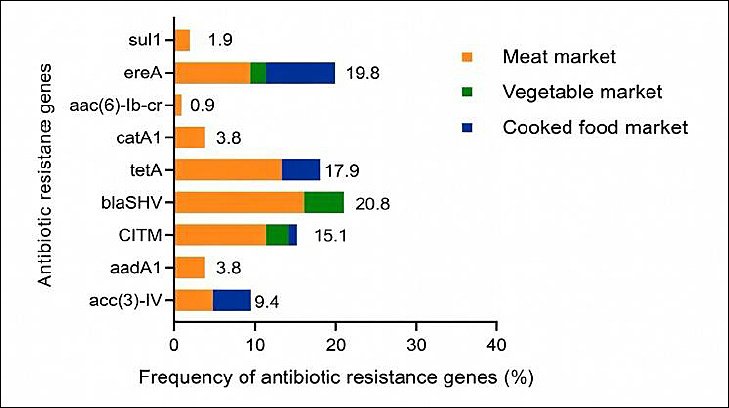
Figure 1. Resistance gene profiles were investigated among Escherichia coli strains obtained from houseflies in different markets. sul1 (sulfonamide), ereA (erythromycin), aac(6)-Ib-cr (ciprofloxacin), catA1 (chloramphenicol), tetA (tetracycline), blaSHV (penicillin), CITM (ampicillin), aadA1 (streptomycin), acc(3)-IV (gentamicin).
Overall, 67 (63.2%) of the 106 isolates carried at least one AMR gene (Table 5). The most frequently detected single gene was blaSHV (12.3%), followed by ereA (11.3%) and CITM (8.5%). Among isolates carrying multiple resistance genes, the most common combinations were acc(3)-IV–tetA (4.7%) and acc(3)-IV–ereA–tetA (4.7%). A significant difference was observed in the distribution of AMR-positive isolates among market types (p = 0.0394).
| Antibiotic resistance gene expression patterns | Meat (n = 79) | Vegetable Market (n = 14) | Cooked-food market (n = 13) | Total (n = 106) | p-value |
|---|---|---|---|---|---|
| Negative | 34 (43.0) | 4 (25.6) | 1 (7.7) | 39 (36.8) | 0.0394 |
| Positive resistance genes | 45 (57.0) | 10 (97.4) | 12 (92.3) | 67 (63.2) | |
| One gene | |||||
| aac(6)-Ib-cr | 1 (1.3) | 0 (0.0) | 0 (0.0) | 1 (0.9) | |
| aadA1 | 2 (2.5) | 0 (0.0) | 0 (0.0) | 2 (1.9) | |
| blaSHV | 8 (10.1) | 5 (35.7) | 0 (0.0) | 13 (12.3) | |
| catA1 | 3 (3.8) | 0 (0.0) | 0 (0.0) | 3 (2.8) | |
| CITM | 5 (6.3) | 3 (21.4) | 1 (7.7) | 9 (8.5) | |
| ereA | 4 (5.1) | 2 (14.3) | 6 (46.2) | 12 (11.3) | |
| tetA | 6 (7.6) | 0 (0.0) | 0 (0.0) | 6 (5.7) | |
| Two genes | |||||
| acc(3)-IV–tetA | 3 (3.8) | 0 (0.0) | 2 (15.4) | 5 (4.7) | |
| blaSHV–CITM | 4 (5.1) | 0 (0.0) | 0 (0.0) | 4 (3.8) | |
| CITM–ereA | 2 (2.5) | 0 (0.0) | 0 (0.0) | 2 (1.9) | |
| Three genes | |||||
| aadA1– blaSHV–tetA | 2 (2.5) | 0 (0.0) | 0 (0.0) | 2 (1.9) | |
| acc(3)-IV–ereA–tetA | 2 (2.5) | 0 (0.0) | 3 (21.3) | 5 (4.7) | |
| blaSHV–CITM–ereA | 1 (1.3) | 0 (0.0) | 0 (0.0) | 1 (0.9) | |
| blaSHV–ereA–sul1 | 1 (1.3) | 0 (0.0) | 0 (0.0) | 1 (0.9) | |
| Four genes | |||||
| blaSHV–catA1–sul1–tetA | 1 (1.3) | 0 (0.0) | 0 (0.0) | 1 (0.9) |
Table 5. Antibiotic resistance gene patterns of Escherichia coli strains obtained from houseflies in different markets in Phayao Province, Northern Thailand.
| Antibiotic resistance gene expression patterns | Meat (n = 79) | Vegetable Market (n = 14) | Cooked-food market (n = 13) | Total (n = 106) | p-value |
|---|---|---|---|---|---|
| Negative | 34 (43.0) | 4 (25.6) | 1 (7.7) | 39 (36.8) | 0.0394 |
| Positive resistance genes | 45 (57.0) | 10 (97.4) | 12 (92.3) | 67 (63.2) | |
| One gene | |||||
| aac(6)-Ib-cr | 1 (1.3) | 0 (0.0) | 0 (0.0) | 1 (0.9) | |
| aadA1 | 2 (2.5) | 0 (0.0) | 0 (0.0) | 2 (1.9) | |
| blaSHV | 8 (10.1) | 5 (35.7) | 0 (0.0) | 13 (12.3) | |
| catA1 | 3 (3.8) | 0 (0.0) | 0 (0.0) | 3 (2.8) | |
| CITM | 5 (6.3) | 3 (21.4) | 1 (7.7) | 9 (8.5) | |
| ereA | 4 (5.1) | 2 (14.3) | 6 (46.2) | 12 (11.3) | |
| tetA | 6 (7.6) | 0 (0.0) | 0 (0.0) | 6 (5.7) | |
| Two genes | |||||
| acc(3)-IV–tetA | 3 (3.8) | 0 (0.0) | 2 (15.4) | 5 (4.7) | |
| blaSHV–CITM | 4 (5.1) | 0 (0.0) | 0 (0.0) | 4 (3.8) | |
| CITM–ereA | 2 (2.5) | 0 (0.0) | 0 (0.0) | 2 (1.9) | |
| Three genes | |||||
| aadA1– blaSHV–tetA | 2 (2.5) | 0 (0.0) | 0 (0.0) | 2 (1.9) | |
| acc(3)-IV–ereA–tetA | 2 (2.5) | 0 (0.0) | 3 (21.3) | 5 (4.7) | |
| blaSHV–CITM–ereA | 1 (1.3) | 0 (0.0) | 0 (0.0) | 1 (0.9) | |
| blaSHV–ereA–sul1 | 1 (1.3) | 0 (0.0) | 0 (0.0) | 1 (0.9) | |
| Four genes | |||||
| blaSHV–catA1–sul1–tetA | 1 (1.3) | 0 (0.0) | 0 (0.0) | 1 (0.9) |
n = Number of E. coli isolates, % = Percentage within each market group, aac(6)-Ib-cr = Ciprofloxacin resistance gene, aadA1 = Streptomycin resistance gene, acc(3)-IV = Gentamicin resistance gene, blaSHV = Penicillin resistance gene, catA1 = Chloramphenicol resistance gene, CITM = Ampicillin resistance gene, ereA = Erythromycin resistance gene, sul1 = Sulfonamide resistance gene, tetA = Tetracycline resistance gene.
p-values were calculated using the chi-square test for comparisons of AMR gene-positive E. coli isolate distribution among market types.
DISCUSSION
Role of houseflies in pathogen transmission
Houseflies (M. domestica) are among the most common synanthropic insects coexisting with human communities, where they cause nuisance and contribute to public health problems. High housefly densities are often indicative of poor household and environmental hygiene [21]. Houseflies possess a strong capacity to acquire and disseminate pathogens from septic sources to other environments through contamination of their body surfaces, including feet, wings, and mouthparts, thereby acting as mechanical vectors for pathogenic agents and AMR dissemination [22].
Prevalence of E. coli in market-associated houseflies
In this study, E. coli was isolated from houseflies collected from meat, vegetable, and cooked-food markets in Muang District, Phayao Province, Northern Thailand. The overall prevalence of E. coli was 30.3%, with the highest prevalence observed in meat markets (39.5%). These findings are consistent with reports from India, where a comparable prevalence of 30% was observed [23], but higher than that reported from Bangladesh (19%) [24]. In contrast, substantially higher contamination rates ranging from 51.4% to 77% have been documented in other geographic regions [8, 10, 25]. The elevated prevalence observed in meat markets may be attributed to inadequate sanitation and frequent exposure of flies to animal feces and contaminated materials during meat handling and processing [26, 27].
Phylogenetic distribution of E. coli isolates
Phylogenetic analysis revealed that phylogroup A predominated among the E. coli isolates (49.1%), consistent with previous studies identifying phylogroup A as the most prevalent group across diverse sources [28]. This phylogroup is commonly associated with commensal as well as pathogenic E. coli strains in humans and animals. However, the proportion observed in the present study was slightly higher than the approximately 40% prevalence reported in similar investigations conducted in other regions [29].
Virulence gene carriage and pathogenic potential
Virulence gene detection is essential for assessing the pathogenic potential of E. coli, particularly diarrheagenic strains [30]. In the present study, 69.8% of isolates carried at least one virulence gene, indicating substantial pathogenic potential among housefly-associated E. coli. The most frequently detected gene was bfpA (26.4%), which occurred predominantly in isolates from meat markets. This prevalence exceeds that reported from surface water in Australia (24%) [31]. The bfpA gene is a hallmark of EPEC and encodes bundle-forming pili that mediate adherence to intestinal epithelial cells, leading to diarrheal disease [32].
The stx1 gene, associated with EHEC, was detected in 7.5% of isolates, a prevalence slightly lower than that reported by Sobur et al. [33]. The absence of invE and elt, which are associated with EIEC and ETEC, respectively [34], may reflect the low circulation of these pathotypes in the studied setting. The lack of significant differences in virulence gene distribution among market types (p = 0.057) highlights the need for continued surveillance of DEC.
Phylogenetic spread of diarrheagenic virulence markers
The detection of multiple DEC-associated virulence markers (eae, bfpA, aggR, astA, stx1, esth, and estp) across different phylogenetic groups, with bfpA predominating, suggests that these virulence traits are not restricted to specific phylogroups. This distribution aligns with previous observations of genetic heterogeneity among atypical EPEC strains and supports the concept that DEC virulence determinants can spread via horizontal gene transfer, thereby weakening strict phylogroup–virulence associations [34–36].
Antimicrobial resistance patterns in E. coli isolates
The detection of antibiotic-resistant E. coli in houseflies represents a significant public health concern. All isolates exhibited complete resistance to penicillin G and erythromycin, consistent with previous reports documenting high resistance rates to these antimicrobials among housefly-associated E. coli [8, 10, 37, 38]. Elevated resistance to ampicillin (66.0%), tetracycline (35.8%), and chloramphenicol (17.0%) was observed, exceeding rates reported from Bangladesh, Iran, and Nigeria [10, 22, 39]. Conversely, the absence of resistance to imipenem and meropenem corroborates findings from China, indicating sustained efficacy of carbapenems against E. coli [25].
Multidrug resistance and phylogenetic associations
MDR was detected in 46.2% of isolates, with a significantly higher prevalence in meat markets than in other market types, reflecting environmental contamination and antimicrobial exposure [40]. Comparable MDR rates have been reported in urban environments [38], whereas lower rates were observed in dairy farm settings [41]. In contrast, higher MDR prevalence has been documented in hospital-adjacent environments [35]. The most frequent MDR pattern, ampicillin–erythromycin–penicillin–tetracycline, underscores the adaptability of E. coli to commonly used antibiotics [41]. Although no significant association was found between phylogenetic group and MDR phenotype (p = 0.1037), MDR is largely driven by mobile genetic elements rather than phylogenetic background alone [42–44].
Antimicrobial resistance gene profiles and public health implications
AMR gene carriage was detected in 63.2% of isolates, highlighting the role of houseflies as environmental reservoirs of resistance determinants. The most prevalent gene was blaSHV (20.8%), although this rate was lower than those reported in food centers and fish markets [10, 40]. Moderate prevalence of ereA, tetA, and CITM further supports the linkage between phenotypic resistance and underlying genetic mechanisms. Variability in gene prevalence across studies likely reflects geographic and environmental differences [41, 45]. These findings emphasize the importance of sustained surveillance and targeted interventions to limit environmental dissemination of AMR genes.
CONCLUSION
This study demonstrated that E. coli is commonly carried by M. domestica in fresh-food market environments, with an overall prevalence of 30.3%. The highest contamination and MDR burden were consistently observed in meat markets. Phylogroup A predominated, while phylogroup D contributed disproportionately to MDR positivity. A high proportion of isolates (69.8%) harbored at least one DEC-associated virulence gene, with bfpA being the most prevalent, indicating a notable presence of EPEC-linked determinants. Phenotypic resistance was widespread, with universal resistance to penicillin G and erythromycin and an overall MDR prevalence of 46.2%. Moreover, 63.2% of isolates carried at least one AMR gene, with blaSHV, ereA, and tetA being the most frequently detected.
These findings highlight fresh-food markets, particularly meat markets, as critical interfaces for environmental dissemination of virulent and MDR E. coli. The detection of DEC-associated virulence markers and AMR genes in houseflies underscores the importance of incorporating insect vectors into routine AMR surveillance programs. Improved market hygiene, effective waste management, and fly control strategies are essential to reduce the risk of food contamination and subsequent human exposure within a One Health framework.
A key strength of this study lies in its integrated approach, combining prevalence estimation, phylogenetic grouping, virulence profiling, phenotypic resistance testing, and AMR gene detection within the same isolates. The comparison across different market types provides context-specific insights into exposure risk, while the focus on houseflies expands current understanding of non-clinical AMR reservoirs.
This cross-sectional study was confined to a single province, which may limit extrapolation to other regions. The absence of whole-genome sequencing restricted resolution of clonal relationships and mobile genetic elements. In addition, antimicrobial susceptibility was assessed using disc diffusion only, without MIC determination, and direct fly-to-food transmission and human exposure were not quantitatively evaluated.
Future studies should incorporate longitudinal sampling, whole-genome sequencing, and quantitative risk assessment to better elucidate transmission pathways of MDR E. coli. Integrating entomological surveillance with food safety and public health monitoring will strengthen One Health–based AMR control strategies. Evaluating the effectiveness of targeted hygiene and vector control interventions in market settings is also warranted.
Overall, this study provides baseline evidence that M. domestica in fresh-food markets act as important environmental reservoirs of virulent and MDR E. coli. Addressing AMR at the food–environment–human interface requires coordinated surveillance and intervention strategies, reinforcing the critical role of environmental vectors in AMR epidemiology.
DATA AVAILABILITY
The supplementary data can be made available from the corresponding author upon request.
AUTHORS’ CONTRIBUTIONS
KK: Conceptualized and designed the study, collected samples, and performed the experiments. KK, SU, AS, and NS: Analyzed the data and drafted the manuscript. KK, SU, and NS: Reviewed and edited the manuscript. KK also secured funding for the study. OJ and CT: Data analysis. All authors have reviewed and approved the final manuscript of the manuscript.
COMPETING INTERESTS
The authors declare that they have no competing interests.
PUBLISHER’S NOTE
Veterinary World remains neutral with regard to jurisdictional claims in the published institutional affiliations.
ACKNOWLEDGMENTS
This research was supported by the University of Phayao and the Thailand Science Research and Innovation Fund (Fundamental Fund 2026, Grant No. 2260/2568 and FF65-RIM111).
REFERENCES
- Ajulo S, Awosile B. Global antimicrobial resistance and use surveillance system (GLASS 2022):investigating the relationship between antimicrobial resistance and antimicrobial consumption data across the participating countries. PLoS One 2024;19(2):e0297921. [Google Scholar] | [Crossref]
- Hariyanto H, Yahya CQ, Cucunawangsih C, Pertiwi CLP. Antimicrobial resistance and mortality. Afr J Infect Dis 2022;16(2):13-20. [Google Scholar] | [Crossref]
- Ahmed SK, Hussein S, Qurbani K, Ibrahim RH, Fareeq A, Kochr Mahmood KA. Antimicrobial resistance:impacts, challenges, and future prospects. J Med Sci Sur Public Health 2024;2:100081. [Google Scholar] | [Crossref]
- Uddin TM, Chakraborty AJ, Khusro A, Zidan BRM, Mitra S, Emran TB. Antibiotic resistance in microbes:history, mechanisms, therapeutic strategies and future prospects. J Infect Public Health 2021;14(12):1750-1766. [Google Scholar] | [Crossref]
- Bertagnolio S, Dobreva Z, Centner CM, Olaru ID, Donà D, Burzo S. WHO global research priorities for antimicrobial resistance in human health. Lancet Microbe 2024;5(11):100902. [Google Scholar] | [Crossref]
- Pakbin B, Brück WM, Rossen JWA. Virulence factors of enteric pathogenic Escherichia coli :a review. Int J Mol Sci 2021;22:9922. [Google Scholar] | [Crossref]
- Siriphap A, Suwancharoen C, Laenoi W, Kraivuttinun P, Suthienkul O, Prapasawat W. First study on virulence genes, antimicrobial resistance, and integrons in Escherichia coli isolated from cage, free-range, and organic commercial eggs in Phayao Province, Thailand. Vet World 2022;15(9):2293-2301. [Google Scholar] | [Crossref]
- Akter S, Sabuj AAM, Haque ZF, Kafi MA, Rahman MT, Saha S. Detection of antibiotic-resistant bacteria and their resistance genes from houseflies. Vet World 2020;13(2):266-274. [Google Scholar] | [Crossref]
- Park R, Dzialo MC, Spaepen S, Nsabimana D, Gielens K, Devriese H. Microbial communities of the house fly Musca domestica vary with geographical location and habitat. Microbiome 2019;7:147. [Google Scholar] | [Crossref]
- Sobur A, Haque ZF, Sabuj AA, Ievy S, Rahman AT, El Zowalaty ME. Molecular detection of multidrug and colistin-resistant Escherichia coli isolated from house flies in various environmental settings. Future Microbiol 2019;14:847-858. [Google Scholar] | [Crossref]
- Ranjbar R, Izadi M, Hafshejani TT, Khamesipour F. Molecular detection and antimicrobial resistance of Klebsiella pneumoniae from house flies (Musca domestica) in kitchens, farms, hospitals and slaughterhouses. J Infect Public Health 2016;9(4):499-505. [Google Scholar] | [Crossref]
- Kababian M, Mozaffari E, Akbarzadeh K, Kordshouli RS, Saghafipour A, Shams S. Identification of bacteria contaminating Musca domestica (Diptera:Muscidae) collected from animal husbandries. Shiraz E-Med J 2020;21(4):e92018. [Google Scholar] | [Crossref]
- Hossain M, Rahman W, Ali M, Sultana T, Hossain K. Identification and antibiogram assay of Escherichia coli isolated from chicken eggs. J Bio-Sci 2021;29:123-133. [Google Scholar] | [Crossref]
- Dashti AA, Jadaon MM, Abdulsamad AM, Dashti HM. Heat treatment of bacteria:a simple method of DNA extraction for molecular techniques. Kuwait Med J 2009;41(2):117-122. [Google Scholar] | [Crossref]
- Heijnen L, Medema G. Quantitative detection of E coli, E. coli O157 and other shiga toxin producing E. coli in water samples using a culture method combined with real-time PCR. J Water Health 2006;4(4):487-498. [Google Scholar] | [Crossref]
- Clermont O, Bonacorsi S, Bingen E. Rapid and simple determination of Escherichia coli phylogenetic group. Appl Environ Microbiol 2000;66(10):4555-4558. [Google Scholar] | [Crossref]
- Performance standards for antimicrobial susceptibility testing:twenty-third informational supplement. Wayne, PA: Clinical and Laboratory Standards Institute; 2018. [Google Scholar]
- Jain P, Bepari AK, Sen PK, Rafe T, Imtiaz R, Reza HM. High prevalence of multiple antibiotic resistance in clinical E coli isolates from Bangladesh and prediction of molecular resistance determinants using WGS of an XDR isolate. Sci Rep 2021;11:22859. [Google Scholar] | [Crossref]
- Van TTH, Chin J, Chapman T, Tran LT, Coloe PJ. Safety of raw meat and shellfish in Vietnam:an analysis of Escherichia coli isolations for antibiotic resistance and virulence genes. Int J Food Microbiol 2008;124(3):217-223. [Google Scholar] | [Crossref]
- Park CH, Robicsek A, Jacoby GA, Sahm DC. Prevalence in the United States of aac(6')-Ib-cr encoding a ciprofloxacin-modifying enzyme. Antimicrob Agents Chemother 2006;50(11):3953-3955. [Google Scholar] | [Crossref]
- Smallegange RC, Den Otter CJ, Takken W, Knols BGJ. Houseflies, annoying and dangerous. Wageningen: Wageningen Academic Publishers; 2007. p. 281-292. [Google Scholar]
- Nazari M, Mehrabi T, Hosseini SM, Alikhani MY. Bacterial contamination of adult house flies (Musca domestica) and sensitivity of these bacteria to various antibiotics, captured from Hamadan city, Iran. J Clin Diagn Res 2017;11((4)):DC04. [Google Scholar] | [Crossref]
- Chandrakar C, Shakya S, Patyal A, Jain A, Ali SL, Mishra OP. ERIC-PCR-based molecular typing of multidrug-resistant Escherichia coli isolated from houseflies (Musca domestica) in the environment of milk and meat shops. Lett Appl Microbiol 2022;75(6):1549-1558. [Google Scholar] | [Crossref]
- Zuhora FT, Hosen MA, Rumi NA, Rahman MS, Alam J, Hasan MH. Molecular characterization of multidrug-resistant bacteria isolated from the external and internal parts of the housefly. J Adv Biotechnol Exp Ther 2023;6(3):597-609. [Google Scholar] | [Crossref]
- Liu Y, Yang Y, Zhao F, Fan X, Zhong W, Qiao D. Multi-drug resistant Gram-negative enteric bacteria isolated from flies at Chengdu Airport, China. Southeast Asian J Trop Med Public Health 2013;44(6):988-996. [Google Scholar] | [Crossref]
- Mersha G, Asrat D, Zewde BM, Kyule M. Occurrence of Escherichia coli O157:H7 in feces, skin and carcasses from sheep and goats in Ethiopia. Lett Appl Microbiol 2010;50(1):71-76. [Google Scholar] | [Crossref]
- Messele YE, Abdi RD, Yalew ST, Tegegne DT, Emeru BA, Weird GM. Molecular determination of antimicrobial resistance in Escherichia coli isolated from raw meat in Addis Ababa and Bishoftu, Ethiopia. Ann Clin Microbiol Antimicrob 2017;16:55. [Google Scholar] | [Crossref]
- Stoppe NC, Silva JS, Carlos C, Sato MIZ, Saraiva AM, Ottoboni LMM. Worldwide phylogenetic group patterns of Escherichia coli from commensal human and wastewater treatment plant isolates. Front Microbiol 2017;8:2512. [Google Scholar] | [Crossref]
- Mosquito S, Pons MJ, Riveros M, Ruiz J, Ochoa TJ. Diarrheagenic Escherichia coli phylogroups are associated with antibiotic resistance and duration of diarrheal episode. Sci World J 2015;2015:610403. [Google Scholar] | [Crossref]
- Robert E, Grippa M, Nikiema DE, Kergoat L, Koudougou H, Auda Y. Environmental determinants of E coli, link with the diarrheal diseases, and indication of vulnerability criteria in tropical West Africa (Kapore, Burkina Faso). PLoS Negl Trop Dis 2021;15(8):e0009634. [Google Scholar] | [Crossref]
- Sidhu JP, Ahmed W, Hodgers L, Toze S. Occurrence of virulence genes associated with diarrheagenic pathotypes in Escherichia coli isolates from surface water. Appl Environ Microbiol 2013;79(1):328-335. [Google Scholar] | [Crossref]
- Nataro JP, Kaper JB. Diarrheagenic Escherichia coli. Clin Microbiol Rev 1998;11(1):142-201. [Google Scholar] | [Crossref]
- Sobur MA, Sabuj AAM, Sarker R, Rahman AMMT, Kabir SML, Rahman MT. Antibiotic-resistant Escherichia coli and Salmonella spp. associated with dairy cattle and farm environment having public health significance. Vet World 2019;12(7):984-993. [Google Scholar] | [Crossref]
- Fujioka M, Otomo Y, Ahsan CR. A novel single-step multiplex polymerase chain reaction assay for the detection of diarrheagenic Escherichia coli. J Microbiol Methods 2013;92(3):289-292. [Google Scholar] | [Crossref]
- Afset JE, Anderssen E, Bruant G, Harel J, Wieler L, Bergh K. Phylogenetic backgrounds and virulence profiles of atypical enteropathogenic Escherichia coli strains from a case-control study using multilocus sequence typing and DNA microarray analysis. J Clin Microbiol 2008;46(7):2280-2290. [Google Scholar] | [Crossref]
- Kaye KS, Gupta V, Mulgirigama A, Joshi AV, Scangarella-Oman NE, Yu K. Antimicrobial resistance trends in urine Escherichia coli isolates from adult and adolescent females in the United States from 2011 to 2019:rising ESBL strains and impact on patient management. Clin Infect Dis 2021;73(11):1992-1999. [Google Scholar] | [Crossref]
- Siriphap A, Kitti T, Khuekankaew A, Boonlao C, Thephinlap C, Thepmalee C. High prevalence of extended-spectrum beta-lactamase-producing Escherichia coli and Klebsiella pneumoniae isolates:a 5-year retrospective study at a tertiary hospital in Northern Thailand. Front Cell Infect Microbiol 2022;12:955774. [Google Scholar] | [Crossref]
- Zuhora FT, Hosen MB, Rumi NA, Rahman MT, Alam J, Hasan MR. Molecular characterization of multidrug-resistant bacteria isolated from the external and internal parts of the housefly. J Adv Biotechnol Exp Ther 2023;6(3):597. [Google Scholar] | [Crossref]
- Odetoyin B, Adeola B, Olaniran O. Frequency and antimicrobial resistance patterns of bacterial species isolated from the body surface of the housefly (Musca domestica) in Akure, Ondo State, Nigeria. J Arthropod Borne Dis 2020;14(1):88-96. [Google Scholar] | [Crossref]
- Songe MM, Hang'ombe BM, Knight-Jones TJ, Grace D. Antimicrobial-resistant enteropathogenic Escherichia coli and Salmonella spp. in houseflies infesting fish in food markets in Zambia. Int J Environ Res Public Health 2017;14(1):21. [Google Scholar] | [Crossref]
- Dos Santos Alves TS, Lara GHB, Maluta RP, Ribeiro MG, Leite DS. Carrier flies of multidrug-resistant Escherichia coli as potential dissemination agent in dairy farm environment. Sci Total Environ 2018;633:1345-1351. [Google Scholar] | [Crossref]
- Mathers AJ, Peirano G, Pitout JD. The role of epidemic resistance plasmids and international high-risk clones in the spread of multidrug-resistant Enterobacteriaceae. Clin Microbiol Rev 2015;28(3):565-591. [Google Scholar] | [Crossref]
- Skurnik D, Lacheeb S, Bernede C, le Menac'h A, Elbaz S, Mohler J. Integrons and antibiotic resistance in phylogenetic group B2 Escherichia coli. Microb Drug Resist 2009;15(3):173-178. [Google Scholar] | [Crossref]
- Petty NK, Ben Zakour NL, Stanton-Cook M, Skippington E, Totsika M, Forde BM. Global dissemination of a multidrug resistant Escherichia coli clone. Proc Natl Acad Sci U S A 2014;111(15):5694-5699. [Google Scholar] | [Crossref]
- Bertelloni F, Bresciani F, Cagnoli G, Scotti B, Lazzerini L, Marcucci M. House flies (Musca domestica) from swine and poultry farms carrying antimicrobial resistant Enterobacteriaceae and Salmonella. Vet Sci 2023;10(2):118. [Google Scholar] | [Crossref]