


ABSTRACT
Background and Aim: Theileria orientalis is an emerging tick-borne hemoprotozoan that has become a major cause of bovine theileriosis, leading to anemia, reduced productivity, and substantial economic losses in cattle. Despite increasing reports from neighboring countries, molecular epidemiological information and phylogenetic evidence for T. orientalis in Iraq remain scarce. This study aimed to determine the molecular prevalence of T. orientalis in cattle and their naturally infesting ticks in Wasit Province, Iraq; identify host-associated risk factors; and characterize the genetic diversity and phylogenetic relationships of local isolates relative to global reference strains.
Materials and Methods: A cross-sectional study was conducted on 170 naturally tick-infested cattle from Wasit Province, Iraq, during July-August 2025. Whole blood and corresponding tick samples were collected from each animal. Genomic DNA was extracted and screened for T. orientalis using conventional polymerase chain reaction targeting the 18S rRNA gene. Positive amplicons were sequenced by Sanger sequencing, followed by sequence alignment, GenBank submission, and phylogenetic analysis using MEGA 11. Associations between infection and animal-related risk factors were evaluated using Chi-square analysis, odds ratio, relative risk, and 95% confidence intervals.
Results: Molecular analysis detected T. orientalis DNA in 21/170 (12.35%) cattle and 16/170 (9.41%) tick samples. Infection prevalence differed significantly according to age and sex. Cattle aged >3-6 years exhibited the highest infection risk, whereas male cattle showed significantly greater positivity and relative risk than females. Sequence analysis demonstrated high genetic conservation among Iraqi isolates. Phylogenetic reconstruction revealed that all cattle isolates clustered closely with a Turkish T. orientalis isolate (HQ197736.1), whereas tick isolates showed close genetic relationships with Turkish isolates (HQ197736.1 and OR211412.1) and a Chinese isolate (PQ207062.1), suggesting regional genetic connectivity and possible transboundary circulation of T. orientalis lineages.
Conclusion: This study provides the first molecular detection and comprehensive phylogenetic characterization of T. orientalis simultaneously in cattle and associated tick vectors from Wasit Province, Iraq. The findings establish the first molecular evidence linking genetically related cattle and tick isolates in the country and provide an important baseline for understanding the epidemiology of oriental theileriosis. Expanded nationwide molecular surveillance, genotype-specific characterization using additional genetic markers, and integrated tick management programs are warranted to improve disease monitoring and reduce the impact of T. orientalis infection on the Iraqi cattle industry.
Keywords: 18S rRNA, cattle, genetic characterization, Iraq, molecular epidemiology, phylogenetic analysis, Theileria orientalis, tick-borne disease.
INTRODUCTION
Blood parasites of cattle are microscopic pathogens transmitted primarily by ticks and cause substantial morbidity, mortality, and economic losses through reduced productivity, impaired animal health, and, in some cases, the emergence of drug-resistant strains [1, 2]. Among these pathogens, bovine theileriosis is one of the most important tick-borne diseases and is caused by several Theileria species, each exhibiting distinct host specificity, geographic distribution, and pathogenicity [3]. For example, Theileria annulata infects cattle and other susceptible hosts across tropical, subtropical, and Mediterranean regions, causing severe tropical theileriosis with high morbidity and mortality, whereas T. parva is predominantly distributed in sub-Saharan Africa and is responsible for the highly fatal East Coast fever in cattle [4, 5].
T. orientalis has a worldwide distribution and primarily infects cattle, typically causing a mild, non-transforming, and frequently asymptomatic infection. Nevertheless, the parasite has emerged as an important cause of bovine theileriosis because it can induce hemolytic anemia, reduced milk production, decreased weight gain, reproductive losses, and considerable economic damage to the cattle industry [6, 7]. The genus Theileria possesses a complex life cycle involving schizonts in lymphoid cells and piroplasms within erythrocytes of vertebrate hosts, whereas ticks become infected by ingesting parasitized erythrocytes during blood feeding [8, 9]. Within the tick gut, erythrocytes are lysed, releasing piroplasms that differentiate into gametes and subsequently fuse to form zygotes. These zygotes penetrate the gut epithelium, develop into kinetes, migrate to the salivary glands, and undergo sporogony to produce infective sporozoites. During subsequent blood feeding, sporozoites are transmitted through tick saliva into the mammalian host, where they invade leukocytes, develop into schizonts, and eventually produce erythrocytic piroplasms [10–12]. Although acute clinical signs such as pyrexia and anemia may be absent or transient, T. orientalis infections often persist throughout the host's lifetime, with recrudescence occurring during periods of physiological or environmental stress, including lactation, pregnancy, and sudden environmental changes [13, 14].
Accurate diagnosis of T. orientalis infection is essential for effective epidemiological surveillance and disease control. Conventional microscopic examination of Giemsa-stained blood smears has limited sensitivity and specificity, particularly in animals with low parasitemia or chronic infections. Consequently, molecular diagnostic methods, especially polymerase chain reaction (PCR), have become the preferred approach due to their superior sensitivity, specificity, and ability to detect multiple T. orientalis genotypes [15, 16]. Furthermore, DNA sequencing provides valuable information on genetic diversity, evolutionary relationships, and molecular epidemiology, enabling accurate characterization of circulating isolates and comparison with global reference strains [17, 18].
In Iraq, several investigations have documented the occurrence of different Theileria species in cattle and other domestic animals [19–22]. However, molecular information regarding Theileria infections in ticks remains extremely limited, with only a few published studies investigating tick-associated infections [23]. Moreover, molecular confirmation of T. orientalis in Iraqi cattle has been reported only once, from Fallujah City in Al-Anbar Province [24]. To date, no study has simultaneously investigated genetically related T. orientalis isolates in cattle and their associated ticks or examined their phylogenetic relationships with globally circulating strains. Consequently, important knowledge gaps remain regarding the molecular epidemiology, host-vector relationship, genetic diversity, and potential transmission dynamics of T. orientalis in Iraq.
Despite the increasing recognition of T. orientalis as an emerging tick-borne pathogen worldwide, comprehensive molecular epidemiological studies in Iraq remain scarce. Previous investigations have largely focused on detecting Theileria species in cattle, while molecular characterization of parasites circulating in naturally infesting ticks has received little attention. Furthermore, no previous Iraqi study has simultaneously evaluated cattle and corresponding tick isolates using sequence-based phylogenetic analysis to determine their genetic relatedness and relationship with international reference isolates. This lack of integrated host-vector molecular data limits the current understanding of transmission pathways, genetic diversity, and regional dissemination of T. orientalis, thereby hindering the development of evidence-based surveillance and control strategies.
Therefore, this study aimed to determine the molecular prevalence of T. orientalis in naturally infected cattle and their associated ticks from Wasit Province, Iraq, using PCR targeting the 18S rRNA gene. In addition, the study sought to characterize the genetic diversity of the detected isolates through DNA sequencing, evaluate their phylogenetic relationships with globally reported T. orientalis strains, and investigate host-associated risk factors influencing infection. Collectively, these findings provide the first integrated molecular and phylogenetic evidence linking cattle and tick isolates of T. orientalis in Wasit Province and establish an important baseline for future epidemiological surveillance and control of oriental theileriosis in Iraq.
MATERIALS AND METHODS
Ethical approval
The study protocol was reviewed and approved by the Scientific Committee, College of Veterinary Medicine, University of Wasit, Iraq, and the Institutional Animal Care and Use Committee (IACUC), University of Wasit (Approval No. WU.CVM.CVM-PD-1429; approved on June 25, 2025). All procedures involving animals were conducted in accordance with the institutional guidelines for the ethical care and use of animals in research and complied with internationally accepted principles for veterinary research involving animals. Blood and tick sampling were performed by licensed veterinarians using standard aseptic techniques and appropriate animal restraint to minimize pain, distress, and handling stress. Approximately 5 mL of jugular venous blood was collected using sterile, single-use needles into EDTA-anticoagulated tubes, while attached ticks were carefully removed using sterile forceps to avoid unnecessary tissue injury. All sampled animals were returned to their owners immediately after sample collection without any invasive intervention beyond routine veterinary procedures. Verbal informed consent was obtained from the owners of all participating cattle before sample collection. The study did not involve endangered or protected animal species, and no experimental infections, euthanasia, or procedures that caused prolonged discomfort were performed. Laboratory procedures involving potentially infectious materials were conducted in accordance with appropriate biosafety practices to protect personnel and prevent environmental contamination.
Study period and location
This cross-sectional study was conducted from July to August 2025 in Wasit Province, located in east-central Iraq, approximately 172 km southeast of Baghdad (Figure 1). The province extends between longitudes 44°40′ E and latitudes 32°00′ N and 33°50′ N and shares its eastern border with the Islamic Republic of Iran. The northeastern region is characterized by low-relief plains and anticlinal folds that receive drainage from adjacent mountainous areas. Wasit Province supports a substantial cattle population owing to favorable agroecological conditions, including access to transboundary marshlands, abundant water resources, and productive alluvial plains that facilitate livestock production [25–27].
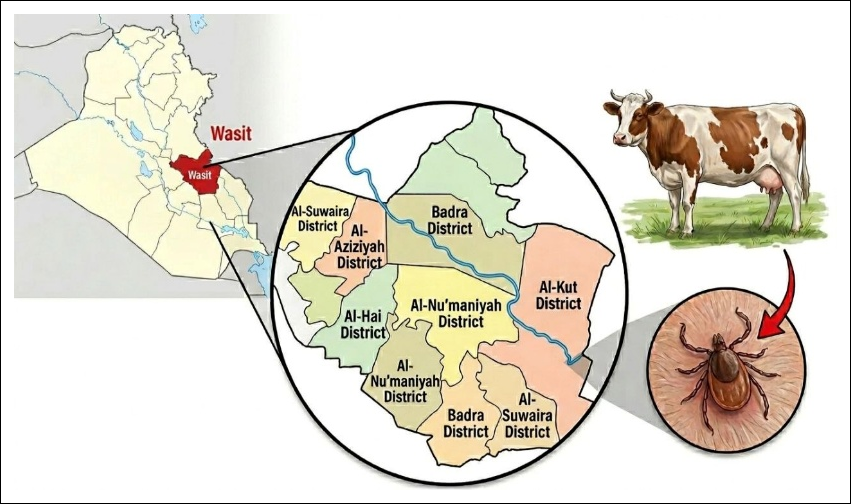
Figure 1. Geographical map of the study areas located in Wasit Province, Iraq.
Study design
A cross-sectional molecular epidemiological study was conducted involving 170 naturally tick-infested crossbred cattle selected from different localities of Wasit Province, Iraq. Animals were enrolled following clinical examination and confirmation of natural tick infestation. All cattle were managed under traditional grazing systems on natural pastures and were frequently exposed to ticks despite routine tick-control practices.
The required sample size was calculated according to the formula described by Thrusfield [28]:
n = Z2Pexp (1-Pexp) / d2
where:
1. n = required sample size;
2. Z = 1.96 (95% confidence interval [CI]);
3. Pexp = expected prevalence (0.50);
4. d = desired precision (0.075).
Sample collection
Under aseptic conditions, approximately 5 mL of jugular venous blood was collected from each animal into ethylenediaminetetraacetic acid (EDTA)-anticoagulated tubes. Hard ticks attached to different body regions of each animal were carefully removed using sterile forceps and placed into individually labeled sterile plastic containers. Blood and tick samples were transported to the laboratory under refrigerated conditions and stored at −20°C until molecular analysis. Information on the age and sex of each animal was recorded for subsequent risk-factor analysis.
Molecular detection of T. orientalis
Genomic DNA was extracted from both blood and tick samples using the gSYNC™ DNA Extraction Kit (Geneaid Biotech, New Taipei City, Taiwan) according to the manufacturer's instructions. DNA concentration and purity were evaluated using a NanoDrop spectrophotometer (Thermo Fisher Scientific, Waltham, MA, USA).
Conventional PCR targeting the 18S rRNA gene was performed using a primer pair designed from the T. orientalis Dunya-30-IRAQ reference sequence (GenBank accession no. OQ779975.1) with Primer3Plus (Wageningen University, Wageningen, the Netherlands). The PCR mixture (20 μL) consisted of 5 μL DNA template, 1 μL forward primer, 1 μL reverse primer, and 13 μL nuclease-free water in AccuPower® PCR PreMix tubes (Bioneer, Daejeon, South Korea).
PCR amplification was carried out using a T100 Thermal Cycler (Bio-Rad Laboratories, Hercules, CA, USA) under the following cycling conditions: initial denaturation at 94°C for 7 min; 30 cycles of denaturation at 94°C for 1 min, annealing at 58°C for 30 s, and extension at 72°C for 1 min; followed by a final extension at 72°C for 7 min.
Amplified PCR products were separated by electrophoresis on 1.5% agarose gels stained with ethidium bromide and electrophoresed at 100 V and 80 mA for 90 min. Positive controls consisted of DNA obtained from recently confirmed Iraqi T. orientalis isolates [24], whereas nuclease-free water served as the negative control. Amplified products were visualized under an ultraviolet transilluminator, and the expected amplicon size was 505 bp.
DNA sequencing and phylogenetic analysis
PCR-positive DNA samples from both cattle and tick specimens, together with the corresponding primers, were submitted to Macrogen Inc. (Seoul, South Korea) for bidirectional DNA sequencing using the modified Sanger sequencing method. The obtained nucleotide sequences were edited and aligned using the ClustalW algorithm. Subsequently, representative sequences were submitted to the National Center for Biotechnology Information (NCBI) GenBank database. Phylogenetic relationships and sequence homology analyses were performed using Molecular Evolutionary Genetics Analysis version 11 (MEGA 11).
Statistical analysis
Statistical analyses were performed using GraphPad Prism version 8 (GraphPad Software Inc., San Diego, CA, USA). The Chi-square (χ²) test was used to evaluate differences among categorical variables. Statistical significance was defined as p < 0.05, with significance levels indicated as follows: p < 0.05 (*), p < 0.01 (**), p < 0.001 (***), and p < 0.0001 (****).
The odds ratio (OR), relative risk (RR), number needed to treat (NNT), and 95% CI were calculated using MedCalc Statistical Software version 22 (MedCalc Software Ltd., Ostend, Belgium) [29].
RESULTS
Incidence of tick infestation according to age and sex
Based on clinical examination, the incidence and risk of tick infestation varied significantly according to the age and sex of the study cattle (Table 1). The incidence of tick infestation differed significantly among age groups (p = 0.0149). Cattle aged >3–6 years exhibited the highest infestation rate (36.47%) and RR (0.9388; p = 0.0008), whereas cattle aged <1 year showed the lowest infestation rate (13.53%) and RR (0.2918). Intermediate infestation rates were observed in cattle aged 1–3 years (28.82%) and >6 years (21.18%).
Sex was also significantly associated with tick infestation. Female cattle exhibited a significantly higher infestation rate than males (84.71% vs. 15.29%; p = 0.0039). Likewise, the RR of tick infestation was markedly higher in females (6.3855; p = 0.0001) than in males (0.3335).
| Factor | No. (%) | RR | NNT | 95% CI |
|---|---|---|---|---|
| Age (years) | ||||
| <1 | 23 (13.53%) | 0.2918 | 3.045 (Benefit) | 2.425 (Benefit) to ∞ to 4.089 (Benefit) |
| 1–3 | 49 (28.82%) | 0.6932 | 7.839 (Benefit) | 4.585 (Benefit) to ∞ to 27.004 (Benefit) |
| >3–6 | 62 (36.47%)* | 0.9388*** | 42.046 (Benefit) | 14.537 (Harm) to ∞ to 8.594 (Benefit) |
| >6 | 36 (21.18%) | 0.4804 | 4.366 (Benefit) | 3.158 (Benefit) to ∞ to 7.073 (Benefit) |
| p-value | 0.0149 | 0.0008 | – | – |
| Sex | ||||
| Female | 144 (84.71%)** | 6.3855**** | 1.4 (Harm) | 1.556 (Harm) to ∞ to 1.272 (Harm) |
| Male | 26 (15.29%) | 0.3335 | 3.272 (Benefit) | 2.561 (Benefit) to ∞ to 4.528 (Benefit) |
| p-value | 0.0039 | 0.0001 | – | – |
Table 1. Incidence and risk of tick infestation according to the age and sex of the study cattle (n = 170).
| Factor | No. (%) | RR | NNT | 95% CI |
|---|---|---|---|---|
| Age (years) | ||||
| <1 | 23 (13.53%) | 0.2918 | 3.045 (Benefit) | 2.425 (Benefit) to ∞ to 4.089 (Benefit) |
| 1–3 | 49 (28.82%) | 0.6932 | 7.839 (Benefit) | 4.585 (Benefit) to ∞ to 27.004 (Benefit) |
| >3–6 | 62 (36.47%)* | 0.9388*** | 42.046 (Benefit) | 14.537 (Harm) to ∞ to 8.594 (Benefit) |
| >6 | 36 (21.18%) | 0.4804 | 4.366 (Benefit) | 3.158 (Benefit) to ∞ to 7.073 (Benefit) |
| p-value | 0.0149 | 0.0008 | – | – |
| Sex | ||||
| Female | 144 (84.71%)** | 6.3855**** | 1.4 (Harm) | 1.556 (Harm) to ∞ to 1.272 (Harm) |
| Male | 26 (15.29%) | 0.3335 | 3.272 (Benefit) | 2.561 (Benefit) to ∞ to 4.528 (Benefit) |
| p-value | 0.0039 | 0.0001 | – | – |
RR = Relative risk; NNT = Number needed to treat; CI = Confidence interval.
Molecular detection of T. orientalis
Conventional PCR detected T. orientalis DNA in 21 of 170 (12.35%) cattle blood samples and 16 of 170 (9.41%) corresponding tick samples (Figure 2).
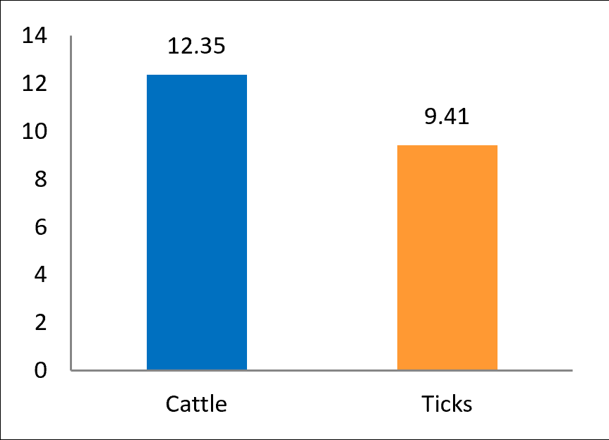
Figure 2. Molecular detection of Theileria orientalis infection among cattle and their naturally infesting ticks by polymerase chain reaction.
Distribution of T. orientalis infection according to host-related risk factors
The prevalence of T. orientalis infection differed significantly among age groups (p = 0.043) (Table 2). The highest prevalence was observed in cattle aged 1–3 years (14.29%) and >3–6 years (14.52%), whereas lower prevalence was observed in cattle aged <1 year (8.70%) and >6 years (8.33%).
Similarly, cattle aged >3–6 years exhibited the highest OR (1.5376) and RR (1.2676), whereas animals aged >6 years showed the lowest OR (0.5859) and RR (0.6496). These findings indicate that cattle aged >3 to 6 years had the greatest likelihood of T. orientalis infection.
According to sex, male cattle exhibited a significantly higher prevalence of T. orientalis infection than females (19.23% vs. 11.11%; p = 0.0445). Likewise, the OR (1.9048) and RR (1.6129) were significantly greater in males than females (p = 0.0001).
Genetic characterization and phylogenetic analysis of T. orientalis
Sequencing analysis of the detected T. orientalis isolates resulted in the submission of nucleotide sequences to the NCBI GenBank database under the isolate designations IS.HA.C1–IS.HA.C21 for cattle isolates and IS.HA.T1–IS.HA.T16 for tick isolates. The corresponding accession numbers ranged from PX851761.1 to PX851781.1 for cattle isolates and from PX851782.1 to PX851797.1 for tick isolates.
Comparative sequence analysis showed nucleotide identities of 78.76% to 100% for cattle isolates and 98.16% to 100% for tick isolates, with sequence variation of 0.001% to 0.05%. Phylogenetic analyses performed separately for cattle and tick isolates revealed that all cattle-derived T. orientalis isolates clustered closely with the Turkish reference isolate HQ197736.1. In contrast, tick-derived isolates exhibited the highest sequence identity with one Chinese isolate (PQ207062.1) and two Turkish isolates (OR211412.1 and HQ197736.1), suggesting close genetic relationships between Iraqi isolates and geographically neighboring T. orientalis strains (Tables 3 and 4; Figures 3–6).
| Factor | Total no. | Positive, no. (%) | OR | RR |
|---|---|---|---|---|
| Age (years) | ||||
| <1 | 23 | 2 (8.70%) | 0.6416 | 0.6989 |
| 1–3 | 49 | 7 (14.29%)* | 1.2738 | 1.2054 |
| >3–6 | 62 | 9 (14.52%)* | 1.5376* | 1.2676* |
| >6 | 36 | 3 (8.33%) | 0.5859 | 0.6496 |
| p-value | – | 0.043 | 0.0001 | 0.0001 |
| 95% CI | – | 6.042–16.88 | 0.2614–17.58 | 0.4363–14.74 |
| Sex | ||||
| Female | 144 | 16 (11.11%) | 0.5250 | 0.6200 |
| Male | 26 | 5 (19.23%)* | 1.9048**** | 1.6129**** |
| p-value | – | 0.0445 | 0.0001 | 0.0001 |
| 95% CI | – | 36.42–66.76 | 7.551–99.81 | 5.192–74.24 |
Table 2. Distribution of Theileria orientalis infection according to host-related risk factors.
| Factor | Total no. | Positive, no. (%) | OR | RR |
|---|---|---|---|---|
| Age (years) | ||||
| <1 | 23 | 2 (8.70%) | 0.6416 | 0.6989 |
| 1–3 | 49 | 7 (14.29%)* | 1.2738 | 1.2054 |
| >3–6 | 62 | 9 (14.52%)* | 1.5376* | 1.2676* |
| >6 | 36 | 3 (8.33%) | 0.5859 | 0.6496 |
| p-value | – | 0.043 | 0.0001 | 0.0001 |
| 95% CI | – | 6.042–16.88 | 0.2614–17.58 | 0.4363–14.74 |
| Sex | ||||
| Female | 144 | 16 (11.11%) | 0.5250 | 0.6200 |
| Male | 26 | 5 (19.23%)* | 1.9048**** | 1.6129**** |
| p-value | – | 0.0445 | 0.0001 | 0.0001 |
| 95% CI | – | 36.42–66.76 | 7.551–99.81 | 5.192–74.24 |
OR = Odds ratio; RR = Relative risk; CI = Confidence interval.
| Local study isolate | Global NCBI-BLAST isolate | ||||||
|---|---|---|---|---|---|---|---|
| Name | Accession no. | Isolate | Accession no. | Host | Country | Identity (%) | |
| IS.HA.C1 | PX851761.1 | Giresun-1 | HQ197736.1 | Bovine | Turkey | 99.75 | |
| IS.HA.C2 | PX851762.1 | Giresun-1 | HQ197736.1 | Bovine | Turkey | 99.69 | |
| IS.HA.C3 | PX851763.1 | Giresun-1 | HQ197736.1 | Bovine | Turkey | 99.73 | |
| IS.HA.C4 | PX851764.1 | Giresun-1 | HQ197736.1 | Bovine | Turkey | 99.81 | |
| IS.HA.C5 | PX851765.1 | Giresun-1 | HQ197736.1 | Bovine | Turkey | 99.24 | |
| IS.HA.C6 | PX851766.1 | Giresun-1 | HQ197736.1 | Bovine | Turkey | 99.59 | |
| IS.HA.C7 | PX851767.1 | Giresun-1 | HQ197736.1 | Bovine | Turkey | 99.62 | |
| IS.HA.C8 | PX851768.1 | Giresun-1 | HQ197736.1 | Bovine | Turkey | 99.71 | |
| IS.HA.C9 | PX851769.1 | Giresun-1 | HQ197736.1 | Bovine | Turkey | 98.76 | |
| IS.HA.C10 | PX851770.1 | Giresun-1 | HQ197736.1 | Bovine | Turkey | 99.25 | |
| IS.HA.C11 | PX851771.1 | Giresun-1 | HQ197736.1 | Bovine | Turkey | 99.18 | |
| IS.HA.C12 | PX851772.1 | Giresun-1 | HQ197736.1 | Bovine | Turkey | 98.99 | |
| IS.HA.C13 | PX851773.1 | Giresun-1 | HQ197736.1 | Bovine | Turkey | 99.45 | |
| IS.HA.C14 | PX851774.1 | Giresun-1 | HQ197736.1 | Bovine | Turkey | 99.75 | |
| IS.HA.C15 | PX851775.1 | Giresun-1 | HQ197736.1 | Bovine | Turkey | 98.92 | |
| IS.HA.C16 | PX851776.1 | Giresun-1 | HQ197736.1 | Bovine | Turkey | 99.15 | |
| IS.HA.C17 | PX851777.1 | Giresun-1 | HQ197736.1 | Bovine | Turkey | 99.68 | |
| IS.HA.C18 | PX851778.1 | Giresun-1 | HQ197736.1 | Bovine | Turkey | 99.75 | |
| IS.HA.C19 | PX851779.1 | Giresun-1 | HQ197736.1 | Bovine | Turkey | 99.41 | |
| IS.HA.C20 | PX851780.1 | Giresun-1 | HQ197736.1 | Bovine | Turkey | 99.62 | |
| IS.HA.C21 | PX851781.1 | Giresun-1 | HQ197736.1 | Bovine | Turkey | 99.70 | |
Table 3. Homology sequence identity (%) between cattle-derived Theileria orientalis isolates and global NCBI-BLAST reference isolates.
| Local study isolate | Global NCBI-BLAST isolate | ||||||
|---|---|---|---|---|---|---|---|
| Name | Accession no. | Isolate | Accession no. | Host | Country | Identity (%) | |
| IS.HA.C1 | PX851761.1 | Giresun-1 | HQ197736.1 | Bovine | Turkey | 99.75 | |
| IS.HA.C2 | PX851762.1 | Giresun-1 | HQ197736.1 | Bovine | Turkey | 99.69 | |
| IS.HA.C3 | PX851763.1 | Giresun-1 | HQ197736.1 | Bovine | Turkey | 99.73 | |
| IS.HA.C4 | PX851764.1 | Giresun-1 | HQ197736.1 | Bovine | Turkey | 99.81 | |
| IS.HA.C5 | PX851765.1 | Giresun-1 | HQ197736.1 | Bovine | Turkey | 99.24 | |
| IS.HA.C6 | PX851766.1 | Giresun-1 | HQ197736.1 | Bovine | Turkey | 99.59 | |
| IS.HA.C7 | PX851767.1 | Giresun-1 | HQ197736.1 | Bovine | Turkey | 99.62 | |
| IS.HA.C8 | PX851768.1 | Giresun-1 | HQ197736.1 | Bovine | Turkey | 99.71 | |
| IS.HA.C9 | PX851769.1 | Giresun-1 | HQ197736.1 | Bovine | Turkey | 98.76 | |
| IS.HA.C10 | PX851770.1 | Giresun-1 | HQ197736.1 | Bovine | Turkey | 99.25 | |
| IS.HA.C11 | PX851771.1 | Giresun-1 | HQ197736.1 | Bovine | Turkey | 99.18 | |
| IS.HA.C12 | PX851772.1 | Giresun-1 | HQ197736.1 | Bovine | Turkey | 98.99 | |
| IS.HA.C13 | PX851773.1 | Giresun-1 | HQ197736.1 | Bovine | Turkey | 99.45 | |
| IS.HA.C14 | PX851774.1 | Giresun-1 | HQ197736.1 | Bovine | Turkey | 99.75 | |
| IS.HA.C15 | PX851775.1 | Giresun-1 | HQ197736.1 | Bovine | Turkey | 98.92 | |
| IS.HA.C16 | PX851776.1 | Giresun-1 | HQ197736.1 | Bovine | Turkey | 99.15 | |
| IS.HA.C17 | PX851777.1 | Giresun-1 | HQ197736.1 | Bovine | Turkey | 99.68 | |
| IS.HA.C18 | PX851778.1 | Giresun-1 | HQ197736.1 | Bovine | Turkey | 99.75 | |
| IS.HA.C19 | PX851779.1 | Giresun-1 | HQ197736.1 | Bovine | Turkey | 99.41 | |
| IS.HA.C20 | PX851780.1 | Giresun-1 | HQ197736.1 | Bovine | Turkey | 99.62 | |
| IS.HA.C21 | PX851781.1 | Giresun-1 | HQ197736.1 | Bovine | Turkey | 99.70 | |
| Local study isolate | Global NCBI-BLAST isolate | ||||||
|---|---|---|---|---|---|---|---|
| Name | Accession no. | Isolate | Accession no. | Host | Country | Identity (%) | |
| IS.HA.T1 | PX851782.1 | CQMB-cattle-CQ55 | PQ207062.1 | Tick | China | 99.83 | |
| IS.HA.T2 | PX851783.1 | CQMB-cattle-CQ55 | PQ207062.1 | Tick | China | 99.83 | |
| IS.HA.T3 | PX851784.1 | CQMB-cattle-CQ55 | PQ207062.1 | Tick | China | 99.83 | |
| IS.HA.T4 | PX851785.1 | CQMB-cattle-CQ55 | PQ207062.1 | Tick | China | 99.83 | |
| IS.HA.T5 | PX851786.1 | CQMB-cattle-CQ55 | PQ207062.1 | Tick | China | 99.83 | |
| IS.HA.T6 | PX851787.1 | CQMB-cattle-CQ55 | PQ207062.1 | Tick | China | 99.83 | |
| IS.HA.T7 | PX851788.1 | CQMB-cattle-CQ55 | PQ207062.1 | Tick | China | 99.83 | |
| IS.HA.T8 | PX851789.1 | CQMB-cattle-CQ55 | PQ207062.1 | Tick | China | 99.67 | |
| IS.HA.T9 | PX851790.1 | RT4-Sarikamis | OR211412.1 | Cattle | Turkey | 99.73 | |
| IS.HA.T10 | PX851791.1 | Giresun-1 | HQ197736.1 | Bovine | Turkey | 99.64 | |
| IS.HA.T11 | PX851792.1 | CQMB-cattle-CQ55 | PQ207062.1 | Tick | China | 98.16 | |
| IS.HA.T12 | PX851793.1 | CQMB-cattle-CQ55 | PQ207062.1 | Tick | China | 98.87 | |
| IS.HA.T13 | PX851794.1 | CQMB-cattle-CQ55 | PQ207062.1 | Tick | China | 98.82 | |
| IS.HA.T14 | PX851795.1 | CQMB-cattle-CQ55 | PQ207062.1 | Tick | China | 98.97 | |
| IS.HA.T15 | PX851796.1 | CQMB-cattle-CQ55 | PQ207062.1 | Tick | China | 98.86 | |
| IS.HA.T16 | PX851797.1 | CQMB-cattle-CQ55 | PQ207062.1 | Tick | China | 98.82 | |
Table 4. Homology sequence identity (%) between tick-derived T. orientalis isolates and global NCBI-BLAST reference isolates.
| Local study isolate | Global NCBI-BLAST isolate | ||||||
|---|---|---|---|---|---|---|---|
| Name | Accession no. | Isolate | Accession no. | Host | Country | Identity (%) | |
| IS.HA.T1 | PX851782.1 | CQMB-cattle-CQ55 | PQ207062.1 | Tick | China | 99.83 | |
| IS.HA.T2 | PX851783.1 | CQMB-cattle-CQ55 | PQ207062.1 | Tick | China | 99.83 | |
| IS.HA.T3 | PX851784.1 | CQMB-cattle-CQ55 | PQ207062.1 | Tick | China | 99.83 | |
| IS.HA.T4 | PX851785.1 | CQMB-cattle-CQ55 | PQ207062.1 | Tick | China | 99.83 | |
| IS.HA.T5 | PX851786.1 | CQMB-cattle-CQ55 | PQ207062.1 | Tick | China | 99.83 | |
| IS.HA.T6 | PX851787.1 | CQMB-cattle-CQ55 | PQ207062.1 | Tick | China | 99.83 | |
| IS.HA.T7 | PX851788.1 | CQMB-cattle-CQ55 | PQ207062.1 | Tick | China | 99.83 | |
| IS.HA.T8 | PX851789.1 | CQMB-cattle-CQ55 | PQ207062.1 | Tick | China | 99.67 | |
| IS.HA.T9 | PX851790.1 | RT4-Sarikamis | OR211412.1 | Cattle | Turkey | 99.73 | |
| IS.HA.T10 | PX851791.1 | Giresun-1 | HQ197736.1 | Bovine | Turkey | 99.64 | |
| IS.HA.T11 | PX851792.1 | CQMB-cattle-CQ55 | PQ207062.1 | Tick | China | 98.16 | |
| IS.HA.T12 | PX851793.1 | CQMB-cattle-CQ55 | PQ207062.1 | Tick | China | 98.87 | |
| IS.HA.T13 | PX851794.1 | CQMB-cattle-CQ55 | PQ207062.1 | Tick | China | 98.82 | |
| IS.HA.T14 | PX851795.1 | CQMB-cattle-CQ55 | PQ207062.1 | Tick | China | 98.97 | |
| IS.HA.T15 | PX851796.1 | CQMB-cattle-CQ55 | PQ207062.1 | Tick | China | 98.86 | |
| IS.HA.T16 | PX851797.1 | CQMB-cattle-CQ55 | PQ207062.1 | Tick | China | 98.82 | |
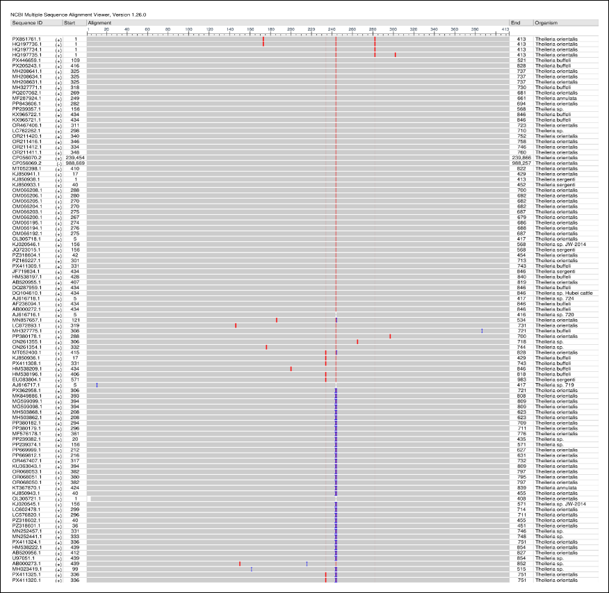
Figure 3. Multiple sequence alignment showing frequency-based nucleotide differences between cattle- and tick-derived Theileria orientalis isolates and NCBI-BLAST reference isolates using the NCBI MSA Viewer.
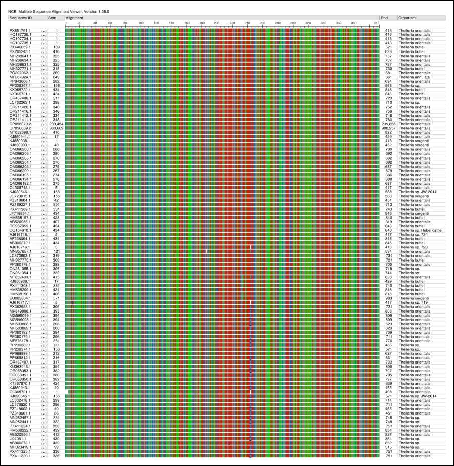
Figure 4. Multiple sequence alignment showing nucleotide sequence alignment of cattle- and tick-derived Theileria orientalis isolates and NCBI-BLAST reference isolates using the NCBI MSA Viewer.
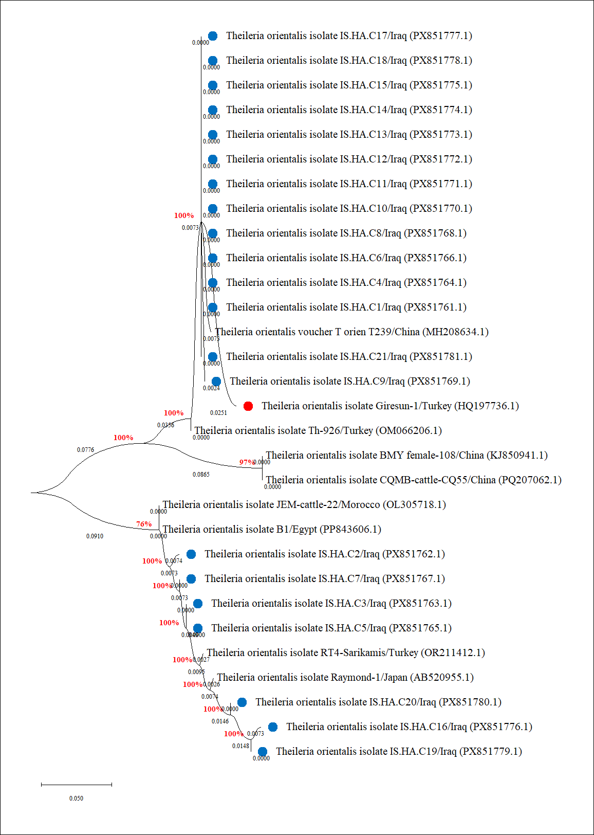
Figure 5. Phylogenetic tree of cattle-derived Theileria orientalis isolates and NCBI-BLAST reference isolates. Study isolates are indicated by blue circles, whereas the closely related Turkish isolate (HQ197736.1) is indicated by a red circle.
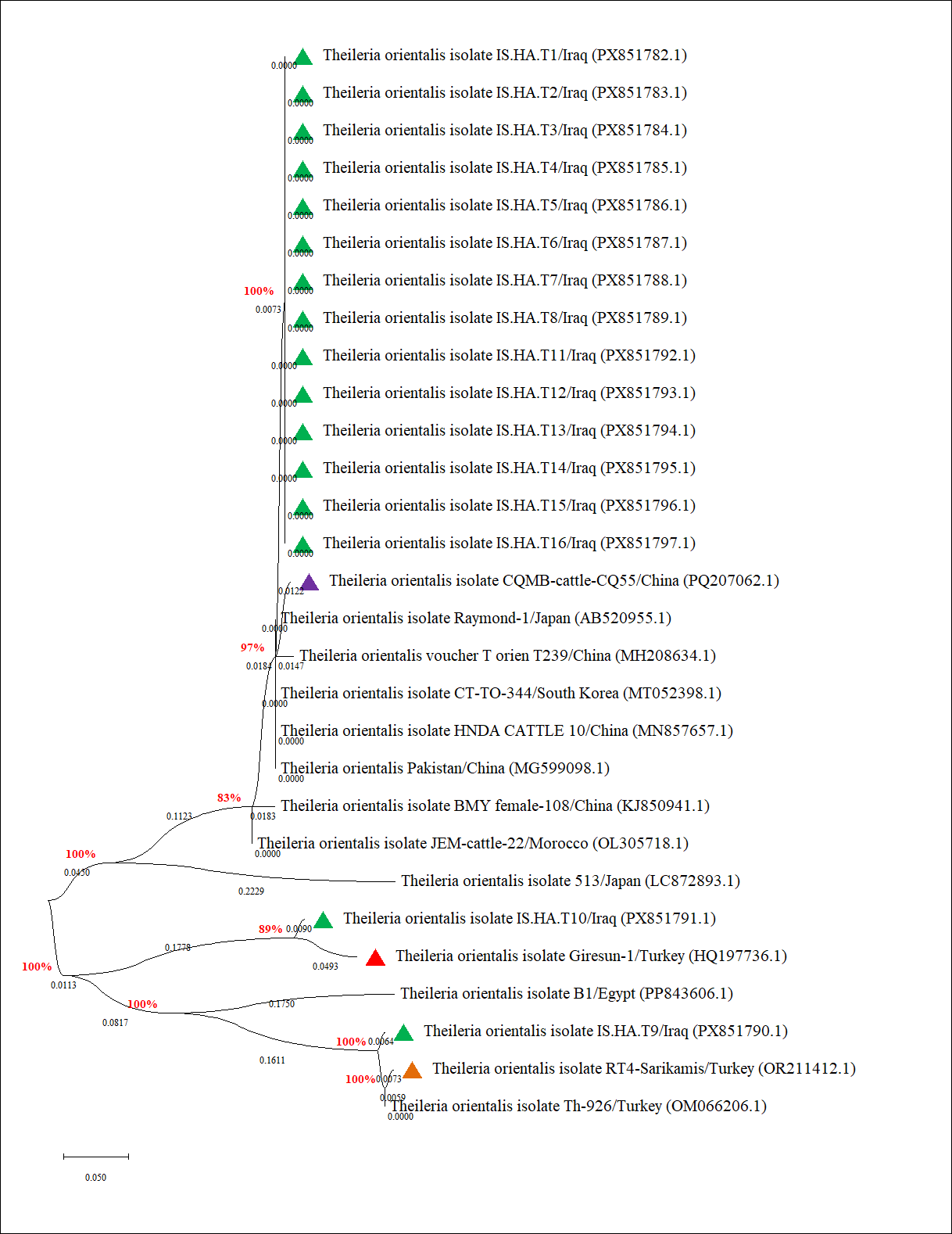
Figure 6. Phylogenetic tree of tick-derived Theileria orientalis isolates and NCBI-BLAST reference isolates. Study isolates are indicated by blue circles, whereas the closely related Turkish (red and brown triangles) and Chinese (purple triangle) reference isolates are highlighted.
DISCUSSION
Molecular prevalence of T. orientalis in cattle and ticks
Information regarding T. orientalis infection in cattle remains limited in Iraq [24], although recent studies conducted in Asia [30, 31], Africa [32, 33], the Americas [34], and Europe [35, 36] have demonstrated that this tick-borne parasite is associated with considerable morbidity, production losses, and, in some cases, mortality in cattle. In the present study, the molecular prevalence of T. orientalis was 12.35% in cattle and 9.41% in their naturally infesting ticks. These findings provide the first simultaneous molecular evidence of T. orientalis infection in cattle and associated ticks from Wasit Province, Iraq.
Compared with previous reports, the prevalence observed in this study was higher than those reported from Ethiopia (2.2%) [37], Turkey (5.6%) [38], and Egypt (8.8%) [39], but was comparable with reports from Vietnam (13.8%) [40] and Pakistan (15.0%) [41]. Lower prevalence than the present study has also been reported in northeastern Thailand (30.1%) [42], Kazakhstan (33.3%) [43], China (14.27%–36.5%) [30, 44], Malaysia (49.76%) [45], Japan (10%–64.8%) [46], and India (83.3%) [31]. Such wide geographical variation in the prevalence of bovine theileriosis is likely attributable to differences in climatic conditions, tick vector abundance, cattle management systems, host immunity, diagnostic approaches, and the genetic diversity of circulating Theileria strains [47].
Environmental factors strongly influence tick ecology, with warm and humid climates generally favoring tick survival and transmission, thereby increasing disease prevalence [48]. In addition, farm management practices, including grazing systems, biosecurity measures, and nutritional status, may alter host susceptibility by increasing physiological stress [49]. Farms managed as small-scale or sideline enterprises often have limited resources for tick control, hygiene, and routine veterinary surveillance, which may further increase the risk of blood parasite transmission [50]. Moreover, genetic variation among Theileria species, including the emergence of strains with different levels of virulence or antimicrobial resistance, may further contribute to regional differences in disease epidemiology [51]. Variability in reported prevalence may also reflect differences in molecular diagnostic procedures, including pre-analytical sample handling, analytical protocols, and biological factors affecting parasite detection.
Association of T. orientalis infection with host-related risk factors
The present study demonstrated that T. orientalis infection was significantly associated with animal age. The highest prevalence was observed in cattle aged 1–3 years and >3–6 years, whereas the highest risk of infection was detected in cattle aged >3–6 years. Likewise, the prevalence of PCR-positive tick samples was greatest among cattle aged 1–3 years. Interpretation of age-related susceptibility should be approached with caution because previous studies have not consistently distinguished between epidemic and endemic disease conditions [6, 52–54].
Previous investigations have reported inconsistent findings regarding the influence of age on T. orientalis infection. In Australia, T. orientalis was detected microscopically in a 4-day-old calf and by quantitative PCR in both newborn and fetal calves [55, 56]. Similarly, Mekata et al. [57] reported that calves born to infected dams were PCR-negative during the first 30 days after birth, whereas 88% became infected by 4 months of age, suggesting that vertical transmission may require several months before becoming detectable under low tick challenge conditions.
In Malaysia, Ola-Fadunsin et al. [45] reported no significant difference in infection prevalence between cattle aged ≤1 year (44.5%) and 1–≤2 years (42.22%), whereas significantly higher prevalence was observed in animals aged 2–≤5 years (52.52%) and >5 years (59.64%). Likewise, Selim et al. [39] demonstrated that asymptomatic cattle older than 3 years had a significantly greater prevalence of T. orientalis infection (13.1%; p < 0.003) than cattle aged <1 year (1.6%) or 1–3 years (5.6%). Conversely, cattle residing in endemic regions, including Iraq, may gradually develop partial protective immunity following repeated exposure to infected ticks, resulting in persistent infection without overt clinical disease [14, 20, 58, 59]. Therefore, higher prevalence in older animals may reflect cumulative exposure rather than increased susceptibility to clinical theileriosis.
Effect of sex on T. orientalis infection
Male cattle exhibited significantly higher infection prevalence, OR, and RR than female cattle. In contrast, the prevalence of T. orientalis infection in ticks was not significantly associated with host sex, although ticks collected from male cattle showed a numerically higher risk of infection.
Previous studies have reported inconsistent associations between sex and T. orientalis infection. Selim et al. [39] found no significant difference between female (9.2%) and male (7.5%) cattle, whereas Ola-Fadunsin et al. [45] reported a higher prevalence among females (51.33%) than males (43.89%). The discrepancy between the present findings and previous reports may reflect differences in herd structure, management practices, and animal utilization. In the present study, most female cattle were maintained for dairy production, whereas male cattle were primarily reared for beef production under more extensive grazing conditions, which may increase their exposure to tick vectors.
Phylogenetic relationships of Iraqi T. orientalis isolates
Although T. orientalis has previously been reported in neighboring countries, including Turkey, Iran, and Pakistan, molecular phylogenetic information from Iraq has been lacking despite the country's strategic geographical position linking Asia and the Mediterranean region. In the present study, phylogenetic analysis demonstrated that all cattle-derived T. orientalis isolates clustered closely with the Turkish isolate HQ197736.1. In contrast, tick-derived isolates exhibited the greatest sequence identity with one Chinese isolate (PQ207062.1) and two Turkish isolates (OR211412.1 and HQ197736.1).
These findings suggest regional genetic connectivity and may reflect transboundary movement of infected animals or tick vectors. The greater genetic diversity observed among tick isolates may be explained by ticks' ability to acquire parasites from multiple infected hosts throughout their life cycle, allowing them to harbor a broader range of T. orientalis genotypes than those circulating within individual cattle populations. Recent outbreaks reported in Asia and Australia have been associated with highly pathogenic genotypes, including the Ikeda genotype, which has expanded into previously unaffected regions [6]. Consequently, the present findings contribute important baseline molecular evidence regarding the circulation and potential dissemination of T. orientalis lineages in Iraq. However, the statement regarding "haplotype diversity and network analysis showing shared versus unique haplotypes between cattle and ticks" should be removed because such analyses were not performed in this study.
Utility of the 18S rRNA gene and study limitations
The 18S rRNA gene remains one of the most reliable molecular markers for detecting T. orientalis because of its high copy number and the presence of conserved regions interspersed with hypervariable domains that enable accurate differentiation from other apicomplexan parasites [60–62]. Furthermore, the conserved nature of the 18S rRNA gene facilitates phylogenetic reconstruction and comparison of Theileria isolates worldwide, providing a robust framework for investigating evolutionary relationships and genotype diversity [63–66].
Nevertheless, several limitations should be acknowledged. The study was limited to a single province in Iraq, which may not fully represent the nationwide epidemiology of T. orientalis. In addition, phylogenetic characterization was based solely on the 18S rRNA gene. More discriminatory genetic markers, such as the MPSP gene, ITS regions, mitochondrial genes, or whole-genome sequencing, were not investigated. Consequently, genotype-specific characterization, identification of polymorphic sites, and differentiation of major genotypes, including Ikeda, Chitose, and Buffeli, were beyond the scope of the present study. Future nationwide molecular surveillance incorporating multiple genetic markers and larger sample sizes is therefore warranted to improve understanding of the genetic diversity, transmission dynamics, and epidemiology of T. orientalis in Iraq.
CONCLUSION
This study provides the first comprehensive molecular evidence of T. orientalis infection in cattle and their naturally infesting ticks from Wasit Province, Iraq, and the first phylogenetic characterization linking host- and vector-derived isolates in the country. Conventional PCR detected T. orientalis in 12.35% of cattle and 9.41% of tick samples. Infection was significantly associated with host age and sex, with cattle aged >3–6 years exhibiting the highest risk of infection, whereas male cattle showed a significantly higher prevalence and risk of infection than females. Phylogenetic analysis based on the 18S rRNA gene demonstrated a high degree of genetic conservation among Iraqi isolates, with cattle isolates clustering closely with a Turkish reference isolate and tick isolates showing close genetic relationships with both Turkish and Chinese isolates, indicating possible regional circulation of genetically related T. orientalis lineages.
From a practical perspective, these findings provide valuable baseline molecular epidemiological data that can support the development of evidence-based surveillance programs, risk-based tick-control strategies, and early molecular diagnosis of oriental theileriosis in Iraq. Simultaneous molecular investigation of cattle and their associated ticks also improves understanding of host-vector relationships and may facilitate more effective integrated control programs to reduce production losses from tick-borne diseases.
A major strength of this study is the combined evaluation of host- and vector-derived samples using molecular detection, DNA sequencing, and phylogenetic analysis, thereby generating the first integrated molecular dataset for T. orientalis in Iraq.
Future studies should include larger nationwide surveys involving different ecological regions, cattle breeds, and tick species while incorporating more discriminatory molecular markers, such as MPSP, ITS, mitochondrial genes, or whole-genome sequencing, to characterize circulating genotypes and clarify transmission dynamics. Longitudinal investigations integrating vector ecology, genotype distribution, and clinical outcomes will further enhance understanding of the epidemiology of T. orientalis and support sustainable control strategies for the Iraqi cattle industry. Overall, this study establishes an important molecular foundation for future surveillance and contributes to the growing body of knowledge on the epidemiology and phylogenetic diversity of T. orientalis in the Middle East.
DATA AVAILABILITY
All data are included within the manuscript.
GENERATIVE AI DECLARATION
The authors declare that no generative artificial intelligence or AI-assisted technologies were used in the writing, analysis, or preparation of this manuscript
AUTHORS’ CONTRIBUTIONS
AHKS: Conceived and designed the study, coordinated field investigations, collected tick samples, and participated in data acquisition. GJKA: Collected blood samples and contributed to field sampling and specimen handling. TKA: Performed molecular laboratory analyses of blood samples. IME: Conducted molecular laboratory analyses of tick samples. ZA: Prepared the initial manuscript draft and contributed to data interpretation. HAJG: Performed statistical analyses, conducted sequence submission to GenBank, carried out phylogenetic and bioinformatics analyses of cattle and tick isolates, supervised the study, and critically revised the manuscript. All authors read, reviewed, and approved the final version of the manuscript.
COMPETING INTERESTS
The authors declare that they have no competing interests.
PUBLISHER’S NOTE
Veterinary World remains neutral with regard to jurisdictional claims in the published institutional affiliations.
ACKNOWLEDGMENTS
The authors sincerely thank all veterinarians who assisted with animal examination, sample collection, and field investigations during this study. Their cooperation and technical support were invaluable to the successful completion of this research. This research received no external funding.
REFERENCES
- de la Fuente J, Estrada-Peña A, Rafael M, Almazán C, Bermúdez S, Abdelbaset AE. Perception of ticks and tick-borne diseases worldwide. Pathogens 2023;12(10):1258. [Google Scholar]
- Strydom T, Lavan RP, Torres S, Heaney K. The economic impact of parasitism from nematodes, trematodes and ticks on beef cattle production. Animals 2023;13(10):1599. [Google Scholar]
- Zeb J, Song B, Aziz MU, Hussain S, Zarin R, Sparagano O. Diversity and distribution of Theileria species and their vectors in ruminants from India, Pakistan and Bangladesh. Diversity 2022;14(2):82. [Google Scholar]
- Kernif T, Medrouh B, Harrat Z, Saidi F, Ziam H. Characterisation of field tropical theileriosis and associated risk factors in two bioclimatic areas of Algeria. Ticks Tick Borne Dis 2024;15(2):102310. [Google Scholar]
- Mukandabvute D, Paul NH, Songwe F, Chipatiko M, Sakwa LL, Chin’ombe N. Theileria parva genetics, prevalence and vaccination practices in Zimbabwe and the African region and the prospects for vaccine development: a systematic review. Vet Res Commun 2025;49(3):146. [Google Scholar]
- Lakew BT, Eastwood S, Walkden-Brown SW. Epidemiology and transmission of Theileria orientalis in Australasia. Pathogens 2023;12(10):1187. [Google Scholar]
- Singh E, Verma S, Sharma D, Parmar D. Insight into oriental theileriosis: Indian perspective. Trop Anim Health Prod 2025;57(9):508. [Google Scholar]
- Clift SJ, Collins NE, Oosthuizen MC, Steyl JC, Lawrence JA, Mitchell EP. The pathology of pathogenic theileriosis in African wild artiodactyls. Vet Pathol 2020;57(1):24-48. [Google Scholar]
- Sojka D, Jalovecká M, Perner J. Babesia, Theileria, Plasmodium and hemoglobin. Microorganisms 2022;10(8):1651. [Google Scholar]
- Sojka D, Franta Z, Horn M, Caffrey CR, Mareš M, Kopáček P. New insights into the machinery of blood digestion by ticks. Trends Parasitol 2013;29(6):276-85. [Google Scholar]
- Kopáček P, Perner J, Sojka D, Šíma R, Hajdušek O. Molecular targets to impair blood meal processing in ticks. In: Ectoparasites: Drug Discovery Against Moving Targets Germany: Wiley-VCH Verlag GmbH & Co. KGaA; 2018. p. 139-65. [Google Scholar]
- Akhtar T, Amanat MU, Wazir N, Naeem MI, Ammar M, Naeem MA. Pathology of parasitic infections. In: Parasitism and Parasitic Control in Animals: Strategies for the Developing World. GB: CABI; 2023. p. 21-39. [Google Scholar]
- Sugimoto C, Fujisaki K. Non-transforming Theileria parasites of ruminants. In: Theileria. Boston, MA: Springer US; 2002. p. 93-106. [Google Scholar]
- Watts JG, Playford MC, Hickey KL. Theileria orientalis: a review. N Z Vet J 2016;64(1):3-9. [Google Scholar]
- Gebrekidan H, Perera PK, Ghafar A, Abbas T, Gasser RB, Jabbar A. An appraisal of oriental theileriosis and the Theileria orientalis complex, with an emphasis on diagnosis and genetic characterisation. Parasitol Res 2020;119(1):11-22. [Google Scholar]
- Sinha S, Kaur U, Sehgal R. Diagnosis of Parasitic Zoonoses. In: Textbook of parasitic zoonoses. Singapore: Springer Nature Singapore; 2022. p. 59-74. [Google Scholar]
- Hilt EE, Ferrieri P. Next generation and other sequencing technologies in diagnostic microbiology and infectious diseases. Genes 2022;13(9):1566. [Google Scholar]
- Gharban HA. First genotyping confirmation of Pichia kudriavzevii in subclinically mastitic cows in Iraq. Rev Ciên Agrovet 2024;23(3):417-24. [Google Scholar]
- Ahmed RA, Mahmmod SL, Kakarash NA, Baba SHekh MO. Conventional and Molecular Diagnosis of Theileriosis (Theileria annulata) in Cattle in Sulaimani Province, Northern Iraq. Passer J Basic Appl Sci 2021;3(2):150-55. [Google Scholar]
- Jassim A, Al-fatlawi MA, Jarad NI, Klaif SF. Clinical and molecular identification of ruling Theileria annulata strains in cattle calves in AL-Diwaniyah province, Iraq. Iraqi J Vet Sci 2021;35(1):115-9. [Google Scholar]
- AlBakri HS, Suleiman EG, Al-Taee AF. Molecular identification of Theileria species in cattle in Mosul city. Iraqi J Vet Sci 2024;38(1):183-9. [Google Scholar]
- Aziz KJ, Hamadamin BQ. Epidemiological and molecular study of Theileria spp. in sheep and goats in Erbil, Iraq. Trop Anim Health Prod 2025;57(2):80. [Google Scholar]
- Al-Fatlawi MSH, Al-Fatlawi MAA. Molecular and phylogenetic study of Theileria spp. isolated from ticks in AL-Diwaniyah city, Iraq. Iraqi J Agric Sci 2019;50(1):475-9. [Google Scholar]
- Salih DAM, Jebur LA, Al-Taii NA, Ibadi WS. Mutation on Theileria species in cattle in Fallujah city, west region of Iraq. Open Vet J 2024;14(9):2261-8. [Google Scholar]
- Al-Abadi AM. Modeling of groundwater productivity in northeastern Wasit Governorate, Iraq using frequency ratio and Shannon’s entropy Models. Appl Water Sci 2017;7(2):699-716. [Google Scholar]
- Al-Badri BH. Financial and economic evaluation of the livestock fund of the agricultural initiative in Iraq for the period (2009-2018). Iraqi J Agric Sci 2021;52(3):647-57. [Google Scholar]
- Shamkhi MS, Al-Badry HJ. Assessment of groundwater recharge potential depending on morphologic analysis in East of Wasit, Southeastern Iraq. Iraqi Geol J 2021;54(2D):138-54. [Google Scholar]
- Thrusfield M. Veterinary Epidemiology Hoboken, NJ: Wiley-Blackwell; 2018. p. 275-372. [Google Scholar]
- Al-Abedi GJ, Sray AH, Hussein AJ, Gharban HA. Molecular detection and blood profiles evaluation of naturally infected camels with subclinical Trypanosoma evansi, Iraq. Ann Trop Med Public Health 2018;23(S20):SP232243. [Google Scholar]
- Chai Y, Che J, Zhao S, Wang J, Guan G, Yin H. Expanding the molecular understanding of Theileria infections in cattle and yaks from Gansu and Qinghai, China. Parasitol Res 2025;124(6):63. [Google Scholar]
- Reghu G, Varghese A, Kalarickal DC, Gopalan AKK, Moosaripparambil ST, Aravindakshan A. Phylogenetic, pathotyping and haplotype analysis of Theileria orientalis in crossbred cattle of northern Kerala, India. 2025. [Google Scholar]
- El-Alfy ES, Abbas I, Baghdadi HB, El-Sayed SAES, Ji S, Rizk MA. Molecular epidemiology and species diversity of tick-borne pathogens of animals in Egypt: a systematic review and meta-analysis. Pathogens 2022;11(8):912. [Google Scholar]
- Kenaw B, Shimelis S, Sibhat B. Molecular characterization of Anaplasma, Babesia, Coxcella, Eherlicha and Theilera pathogens Circulating in Cattle in Assosa and Bambasi Districts, Benishangul Gumuz Region, Western Ethiopia. Dire Dawa: Haramaya University; 2023. [Google Scholar]
- Iduu N, Barua S, Falkenberg S, Armstrong C, Stockler JW, Moye A. Theileria orientalis Ikeda in cattle, Alabama, USA. Vet Sci 2023;10(11):638. [Google Scholar]
- Kovalchuk SN. Molecular characterization and phylogenetic study of Theileria sp. parasites detected in cattle from the Moscow region of Russia. Ticks Tick Borne Dis 2022;13(1):101835. [Google Scholar]
- Stevanović O, Radalj A. Molecular evidence of Theileria orientalis infection in cattle from Bosnia and Herzegovina. Vet Glas 2023;77(1):86-80. [Google Scholar]
- Gebrekidan H, Gasser RB, Baneth G, Yasur-Landau D, Nachum-Biala Y, Hailu A. Molecular characterization of Theileria orientalis from cattle in Ethiopia. Ticks Tick Borne Dis 2016;7(5):742-7. [Google Scholar]
- Zhou M, Cao S, Sevinc F, Sevinc M, Ceylan O, Moumouni PFA. Molecular detection and genetic identification of Babesia bigemina, Theileria annulata, Theileria orientalis and Anaplasma marginale in Turkey. Ticks Tick Borne Dis 2016;7(1):126-34. [Google Scholar]
- Selim A, Attia K, AlKahtani MD, Albohairy FM, Shoulah S. Molecular epidemiology and genetic characterization of Theileria orientalis in cattle. Trop Anim Health Prod 2022;54(3):178. [Google Scholar]
- Khukhuu A, Lan DTB, Long PT, Ueno A, Li Y, Luo Y. Molecular epidemiological survey of Theileria orientalis in Thua Thien Hue province, Vietnam. J Vet Med Sci 2011;73(5):701-5. [Google Scholar]
- Gebrekidan H, Abbas T, Wajid M, Ali A, Gasser RB, Jabbar A. Molecular characterisation of Theileria orientalis in imported and native bovines from Pakistan. Infect Genet Evol 2017;47:19-25. [Google Scholar]
- Jirapattharasate C, Moumouni PFA, Cao S, Iguchi A, Liu M, Wang G. Molecular epidemiology of bovine Babesia spp. and Theileria orientalis parasites in beef cattle from northern and northeastern Thailand. Parasitol Int 2016;65(1):62-9. [Google Scholar]
- Kuibagarov M, Makhamed R, Zhylkibayev A, Berdikulov M, Abdrakhmanov S, Kozhabayev M. Theileria and Babesia infection in cattle–First molecular survey in Kazakhstan. Ticks Tick Borne Dis 2023;14(1):102078. [Google Scholar]
- Wang J, Yang J, Liu J, Wang X, Xu J, Liu A. Molecular detection and genetic diversity of Theileria orientalis in cattle in China. Parasitol Res 2018;117(12):3689-94. [Google Scholar]
- Ola-Fadunsin SD, Sharma RSK, Abdullah DA, Gimba FI, Jesse FFA, Sani RA. Molecular detection, prevalence and risk factors of Theileria orientalis infection among cattle in Peninsular Malaysia. Prev Vet Med 2020;180:105027. [Google Scholar]
- Ota N, Mizuno D, Kuboki N, Igarashi I, Nakamura Y, Yamashina H. Epidemiological survey of Theileria orientalis infection in grazing cattle in the eastern part of Hokkaido, Japan. J Vet Med Sci 2009;71(7):937-44. [Google Scholar]
- Amzati GS, Djikeng A, Odongo DO, Nimpaye H, Sibeko KP, Muhigwa JBB. Genetic and antigenic variation of the bovine tick-borne pathogen Theileria parva in the Great Lakes region of Central Africa. Parasit Vectors 2019;12(1):588. [Google Scholar]
- Gopalakrishnan B, Sugumaran MP, Kannan B, Thirunavukkarasu M, Davamani V. Assessing the influence of biotic and abiotic factors on tick disease incidence in cattle. Adv Anim Vet Sci 2020;8(11):1120-8. [Google Scholar]
- Sahoo S, Sahoo N, Biswal S, Mohanty BN, Behera B, Pahari A. Theileria orientalis Buffeli pathotype in cows in a theileriosis-endemic region of India. Trop Biomed 2023;40(2):236-40. [Google Scholar]
- Ritonga MZ, Putra A, Prastia A, Nasution F, Br Ginting R. Detection Of Blood Parasites In Cattle In Kutalimbaru Subdistrict, Deli Serdang Regency, North Sumatera. E3S Web Conf 2020;151:10-40. [Google Scholar]
- Nehra AK, Kumari A, Moudgil AD, Vohra S. An insight into misidentification of the small-subunit ribosomal RNA (18S rRNA) gene sequences of Theileria spp. as Theileria annulata. BMC Vet Res 2022;18(1):454. [Google Scholar]
- Abdela N, Bekele T. Bovine theileriosis and its control: a review. Adv Biol Res 2016;10(4):200-12. [Google Scholar]
- Kiara H, Steinaa L, Nene V, Svitek N. Theileria in ruminants. In: Parasitic protozoa of farm animals and pets. Cham: Springer International Publishing; 2017. p. 187-213. [Google Scholar]
- Gharbi M, Darghouth MA, Elati K, AL-Hosary AA, Ayadi O, Salih DA. Current status of tropical theileriosis in Northern Africa: A review of recent epidemiological investigations and implications for control. Transbound Emerg Dis 2020;67(Suppl 1):8-25. [Google Scholar]
- Swilks E, Jenkins C, Poynting A, Collins D, Krebs GL. Prevalence and effect of Theileria orientalis infection in homebred calves in the Gloucester region of New South Wales, Australia. Aust Vet J 2017;95(6):211-6. [Google Scholar]
- Swilks E, Fell SA, Hammer JF, Sales N, Krebs GL, Jenkins C. Transplacental transmission of Theileria orientalis occurs at a low rate in field-affected cattle: infection in utero does not appear to be a major cause of abortion. Parasit Vectors 2017;10(1):227. [Google Scholar]
- Mekata H, Minamino T, Mikurino Y, Yamamoto M, Yoshida A, Nonaka N. Evaluation of the natural vertical transmission of Theileria orientalis. Vet Parasitol 2018;263:1-4. [Google Scholar]
- Tawfeeq DA, AlBakri HS. Clinical, microscopical and molecular detection of caprine theileriosis. Iraqi J Vet Sci 2024;38(3):693-9. [Google Scholar]
- Taylor MA, Coop RL, Wall R. Veterinary parasitology. USA: John Wiley & Sons; 2015. [Google Scholar]
- Ullah I, Khan T, Shuaib N, Fawad M, Gohar S. Prevalence Statistics of Anaplasmosis in Cows of District Charsadda, KP, Pakistan: Microscopy and Statistical analysis of Anaplasma. Sciencetech 2024;5(2):63-74. [Google Scholar]
- Mofokeng LS. Molecular characterization of protozoan parasites and Ehrlichia in domestic animals from uMkhanyakude district of KwaZulu-Natal. South Africa: North-West University; 2019. [Google Scholar]
- Bawm S, Sagara R, Kakisaka K, Thu MJ, Hmoon MM, Htun LL. Identification, genetic variation, and structural analysis of 18S rRNA of Theileria orientalis and Theileria velifera-like isolates from Myanmar. Parasitol Int 2021;82:102299. [Google Scholar]
- Ajaj EA, Mohammad HA, Gharban HA. First molecular confirmation of Coenurus cerebralis in sheep and goats with neurological behaviors in Iraq. Vet World 2021;14(6):1420-5. [Google Scholar]
- Kozhabaev M, Kuzerbaeva A, Baizhanov K, Tulemetova S, Nurkhodzhaev N. Study of the dynamics of distribution, seasonality, and degree of infection with bovine theileriosis in the territory of Turkestan Region. Int J Vet Sci 2023;12:382-8. [Google Scholar]
- Alberfkani MI, Swar SO, Almutairi LA, Hasan HK, Ahmed AE, Khalid HM. Molecular Characterization and phylogenetic analysis of 18S rRNA, gp60 and HSP70 genes of Cryptosporidium parvum isolated from cattle owners and cattle using nested PCR. Pak Vet J 2024;44(4):1237-42. [Google Scholar]
- MA Z. Molecular epidemiological investigation of tick-borne haemopathogens in ticks and livestock in Turkey. Obihiro: Obihiro University of Agriculture and Veterinary Medicine; 2025. [Google Scholar]