


ABSTRACT
Background and Aim: The increasing cost and limited availability of fishmeal have intensified the search for sustainable alternative protein sources in aquaculture. Fermentation of plant-based feed ingredients can improve their nutritional value and functional properties. This study evaluated the effects of dietary inclusion of Leucaena leucocephala leaves fermented with Lactiplantibacillus plantarum TISTR1284 as a partial fishmeal substitute on immune responses, intestinal health, and resistance to Aeromonas hydrophila infection in giant freshwater prawns (Macrobrachium rosenbergii).
Materials and Methods: Adult M. rosenbergii were randomly assigned to three dietary treatments: a control diet and diets in which 10% or 20% of fishmeal was replaced with fermented L. leucocephala. Prawns were fed the experimental diets for 2 weeks before challenge with A. hydrophila. Total hemocyte counts, phenoloxidase activity, hematopoietic tissue proliferation, immune-related gene expression, intestinal histology, antioxidant properties of the diets, and post-challenge survival were evaluated.
Results: Fermentation enhanced the nutritional quality of L. leucocephala by increasing essential amino acid content and reducing mimosine concentration. Diets containing fermented L. leucocephala exhibited greater antioxidant activity, total phenolic content, and total flavonoid content than the control diet. Prawns receiving fermented diets showed significantly increased total hemocyte counts, enhanced hematopoietic cell proliferation, elevated crustacean hematopoietic factor expression, and increased phenoloxidase activity. The expression of key immune-related genes, including IMD, Relish, HSP70, Cu/Zn-SOD, and anti-lipopolysaccharide factor (ALF), was upregulated, whereas TRAF6 and Dorsal expression was moderated during infection. Intestinal morphology remained normal, indicating that dietary inclusion of fermented L. leucocephala did not adversely affect gut structure. Following A. hydrophila challenge, prawns fed fermented diets exhibited a 26.14% reduction in mortality compared with the control group, demonstrating improved disease resistance.
Conclusion: Partial replacement of fishmeal with L. leucocephala leaves fermented by L. plantarum TISTR1284 enhanced innate immune responses, improved antioxidant status, and increased resistance to A. hydrophila infection in M. rosenbergii without compromising intestinal health. Fermented L. leucocephala represents a promising sustainable functional feed ingredient for freshwater prawn aquaculture and may contribute to reducing dependence on fishmeal in commercial production systems.
Keywords: Aeromonas hydrophila, disease resistance, fishmeal replacement, giant freshwater prawn, immune response, Lactiplantibacillus plantarum, Leucaena leucocephala, sustainable aquaculture.
INTRODUCTION
Protein is an essential nutrient in aquaculture diets for crustaceans because it directly influences growth performance, immune function, and cellular processes such as tissue repair, renewal, and reproduction [1]. In Macrobrachium rosenbergii, the recommended dietary crude protein level for achieving optimal growth is approximately 35% of the total diet [2]. Fishmeal remains the predominant protein source in commercial aquafeeds because of its balanced amino acid profile, high digestibility, and low content of anti-nutritional factors (ANFs) [3, 4]. However, increasing aquaculture production and the growing demand for fish-derived products have contributed to rising fishmeal costs and concerns regarding the environmental and ecological consequences of overfishing [5, 6]. Consequently, rising price volatility and limited availability of fishmeal have intensified the search for sustainable, cost-effective alternative protein sources for aquaculture feeds [7, 8]. Plant-derived protein ingredients, including soybean, canola, blue-green microalgae, and cottonseed, have been extensively investigated as potential alternatives to fishmeal in sustainable aquaculture systems because of their favorable protein content and availability [3, 4, 9].
Leucaena leucocephala, a leguminous plant belonging to the family Fabaceae, is widely distributed throughout tropical and subtropical regions of Asia, Africa, and the Americas [10]. The plant contains relatively high protein concentrations (25.25%–30.81%) and provides essential amino acids, including lysine, leucine, proline, and serine, making it a potentially valuable feed ingredient [10]. In marine shrimp (Penaeus vannamei), dietary inclusion of 5%–20% L. leucocephala leaf powder did not adversely affect animal health, biochemical composition, or meat quality [9]. Similarly, replacement of fish protein with 25%–50% L. leucocephala leaf meal devoid of mimosine did not negatively affect the health of Nile tilapia [11]. Furthermore, fish fed diets containing 33% L. leucocephala seed powder exhibited enhanced immune responses during infection with Vibrio harveyi and Pseudomonas aeruginosa [12]. Beyond its nutritional value, L. leucocephala contains bioactive phytochemicals, particularly flavonoids, which possess antioxidant and immunomodulatory properties [13]. Flavonoids have been reported to induce erythropoietin gene expression and exhibit strong anti-inflammatory and antioxidant activities [14]. In aquatic animals, these compounds can interact with hemocyte receptors and activate intracellular signaling pathways associated with immune defense mechanisms [15]. Collectively, previous studies have demonstrated that partial replacement of fishmeal with up to 20% L. leucocephala leaf meal can be implemented without causing severe adverse effects on animal health or growth performance [9].
Despite these advantages, the application of plant-derived proteins in aquafeeds remains constrained by the presence of ANFs, including phytates, trypsin inhibitors, lectins, and other compounds that may reduce palatability, impair nutrient utilization, and adversely affect growth and immune performance [16]. Fermentation has emerged as an effective strategy for improving the nutritional quality and functionality of plant materials through biochemical transformation and detoxification [17]. Among the microorganisms used for this purpose, lactic acid bacteria (LAB), including species of Lactiplantibacillus, Aerococcus, and Streptococcus, have received considerable attention because of their safety and ability to enhance nutrient bioavailability [17, 18]. Fermented products are generally recognized as safe, nutritionally enriched, and stable during storage. Controlled fermentation using selected LAB strains has therefore been proposed as a valuable approach for producing high-quality functional feed ingredients [17].
Lactiplantibacillus plantarum is a particularly promising microorganism because of its capacity to biotransform phenolic-rich substrates and generate bioactive compounds with potent antioxidant activity. This bacterium facilitates the conversion of hydroxybenzoic acids, hydrolyzes tannins to gallic acid, and promotes the transformation of hydroxycinnamic acids into biologically active metabolites [18]. In addition to its well-recognized probiotic properties in humans, L. plantarum has demonstrated beneficial effects in aquaculture by enhancing growth performance, improving immune responses, and increasing disease resistance through the production of antimicrobial compounds that suppress pathogenic bacteria [19]. In M. rosenbergii, dietary supplementation with L. plantarum has been shown to improve host-associated microbiota, feed utilization, carcass composition, immune status, and survival following infection with Aeromonas hydrophila [19].
Although the nutritional and immunological benefits of L. leucocephala and L. plantarum have been independently documented, limited information is available regarding the combined application of these two components as a fermented functional feed ingredient in crustacean aquaculture. Previous studies have primarily focused on direct inclusion of L. leucocephala leaf meal or probiotic supplementation alone, whereas the effects of L. leucocephala fermented with L. plantarum on immune modulation, antioxidant status, intestinal health, and disease resistance have not been investigated in M. rosenbergii. Furthermore, the influence of fermented L. leucocephala on hematopoietic activity, phenoloxidase-mediated defense responses, and immune-related gene expression during bacterial infection remains poorly understood. Addressing these knowledge gaps is essential for evaluating the feasibility of fermented plant-based proteins as sustainable alternatives to fishmeal in freshwater prawn production systems.
Therefore, this study was conducted to evaluate the effects of partial replacement of fishmeal with L. leucocephala leaves fermented by L. plantarum TISTR1284 in diets for M. rosenbergii. Specifically, the study investigated the effects of dietary inclusion of fermented L. leucocephala on antioxidant properties of the diets, total hemocyte count, hematopoietic tissue proliferation, phenoloxidase activity, immune-related gene expression, intestinal morphology, and resistance to A. hydrophila infection. The findings provide insight into the potential application of fermented L. leucocephala as a sustainable functional feed ingredient capable of reducing dependence on fishmeal while enhancing immune competence and disease resistance in freshwater prawn aquaculture.
MATERIALS AND METHODS
Ethical approval
All experimental procedures involving giant freshwater prawns (M. rosenbergii) were reviewed and approved by the Laboratory Animals Research Center, University of Phayao, and the Animal Ethics Committee of the University of Phayao, Thailand (Approval No. 63 01 04 010; approved on 4 August 2020). The study was conducted in accordance with institutional guidelines for the care and use of aquatic animals in research and relevant standards for animal welfare, biosafety, and humane experimental practice.
Healthy adult prawns were acclimatized before the experiment and maintained under controlled water quality conditions to minimize stress. Animal handling, dietary intervention, bacterial challenge with A. hydrophila, hemolymph collection, tissue sampling, and survival monitoring were performed only by trained personnel using standardized procedures. The number of prawns used was limited to the minimum required to achieve the study objectives, and the sample size was determined before experimentation using G*Power software. During the challenge experiment, animals were monitored regularly for abnormal behavior, morbidity, and mortality. All efforts were made to reduce pain, distress, and unnecessary handling throughout the study. Moribund prawns and dead animals were promptly removed from the tanks, and biological waste and bacterial cultures were handled and disposed of in accordance with institutional biosafety procedures.
Study period and location
The study was conducted from March to May 2021 at the laboratory and aquaculture research facilities of the University of Phayao, Phayao, Thailand. Healthy adult M. rosenbergii were obtained from a local commercial farm in Chiang Rai, Thailand, and transported to the laboratory for acclimatization before the feeding trial and bacterial challenge experiment.
Study design
This study evaluated the effects of partial replacement of fishmeal with fermented L. leucocephala on immune responses and resistance to A. hydrophila infection in adult M. rosenbergii. Three dietary treatments were prepared: a control diet, a diet in which 10% of fishmeal was replaced with fermented L. leucocephala, and a diet in which 20% of fishmeal was replaced with fermented L. leucocephala. After acclimatization, prawns were randomly allocated to dietary groups and fed for 2 weeks before challenge with A. hydrophila. Hemolymph, hematopoietic tissue, and intestinal samples were collected at predetermined time points for immunological, histological, and molecular analyses. A separate survival experiment was conducted using three independent replicate tanks per treatment group.
Macrobrachium rosenbergii culture
Healthy M. rosenbergii weighing 25–30 g were obtained from a local farm in Chiang Rai, Thailand, and maintained in the laboratory under acclimatized conditions for 7 days before the start of the experiment. Adult prawns were selected because the present study primarily focused on immune status and immune-related responses following dietary supplementation and bacterial challenge. In addition, their larger body size facilitated the collection of hemolymph for immunological and molecular analyses. During acclimatization, prawns were fed a commercial diet three times daily at 07:00, 13:00, and 19:00 h.
Bacterial culture
The A. hydrophila culture and the protocol for determining the median lethal dose in M. rosenbergii were described previously [20]. The stock culture of L. plantarum TISTR1284 was stored in the laboratory at −80°C in de Man, Rogosa, and Sharpe medium (HiMedia Laboratories Pvt. Ltd., Mumbai, India) containing 20% glycerol. The thawed stock culture was cultivated in fresh medium and incubated at 37°C for 48 h. The culture was harvested, and the optical density at 600 nm was adjusted to 0.1 using 0.85% sterile NaCl solution to obtain an inoculum concentration of 1.5 × 10⁸ CFU/mL.
Preparation of fermented L. leucocephala
L. leucocephala leaf tips were collected, washed, and air-dried. Fermentation was performed using L. plantarum TISTR1284 at an inoculum level of 1 × 10⁶ CFU/g plant material following a modified method described previously [21]. The plant material was packed in airtight containers and incubated under anaerobic conditions at room temperature (25°C–30°C) for 30 days. After fermentation, samples were oven-dried and ground into a fine powder for diet formulation. Amino acid composition of fermented and non-fermented samples was analyzed by Central Laboratory (Thailand) Co., Ltd., Bangkok, Thailand, using high-performance liquid chromatography, and the results were expressed as mg/100 g dry weight (Table 1).
| Amino acid | Non-fermented L. leucocephala (mg/100 g) | Fermented L. leucocephala (mg/100 g) |
|---|---|---|
| Toxic non-protein amino acid | ||
| Mimosine | 92.46 | 53.46 |
| Essential amino acids | ||
| Phenylalanine | 2309 | 2973 |
| Histidine | 1658 | 1680 |
| Leucine | 2291 | 2829 |
| Lysine | 5695 | 7092 |
| Methionine | <20 | <20 |
| Valine | 1063 | 1236 |
| Threonine | 248 | 262 |
| Non-essential amino acids | ||
| Aspartic acid | 1026 | 1216 |
| Glutamic acid | 1413 | 1340 |
| Alanine | 678 | 814 |
| Cystine | 246 | 320 |
| Glycine | 479 | 510 |
| Hydroxyproline | <20 | <20 |
| Hydroxylysine | <20 | <20 |
| Proline | 546 | 641 |
| Serine | 383 | 903 |
| Isoleucine | 1183 | 1374 |
| Tyrosine | 2116 | 2152 |
| Tryptophan | 248 | 245 |
Table 1. Amino acid composition of fermented and non-fermented Leucaena leucocephala.
| Amino acid | Non-fermented L. leucocephala (mg/100 g) | Fermented L. leucocephala (mg/100 g) |
|---|---|---|
| Toxic non-protein amino acid | ||
| Mimosine | 92.46 | 53.46 |
| Essential amino acids | ||
| Phenylalanine | 2309 | 2973 |
| Histidine | 1658 | 1680 |
| Leucine | 2291 | 2829 |
| Lysine | 5695 | 7092 |
| Methionine | <20 | <20 |
| Valine | 1063 | 1236 |
| Threonine | 248 | 262 |
| Non-essential amino acids | ||
| Aspartic acid | 1026 | 1216 |
| Glutamic acid | 1413 | 1340 |
| Alanine | 678 | 814 |
| Cystine | 246 | 320 |
| Glycine | 479 | 510 |
| Hydroxyproline | <20 | <20 |
| Hydroxylysine | <20 | <20 |
| Proline | 546 | 641 |
| Serine | 383 | 903 |
| Isoleucine | 1183 | 1374 |
| Tyrosine | 2116 | 2152 |
| Tryptophan | 248 | 245 |
Amino acid composition was determined by Central Laboratory (Thailand) Co., Ltd., Bangkok, Thailand, using the in-house method TE-CH-372 based on the Official Journal of the European Communities, Commission Directive 98/64/EC, Annex Part A, L257 (1998), pp. 14–23. Values are expressed as mg/100 g dry weight.
Antioxidant and phytochemical activities were analyzed as shown in Table 2. Fermentation increased most essential and non-essential amino acids while reducing mimosine content, indicating improved nutritional quality of L. leucocephala leaves (Table 1).
Experimental diets
Three diets were prepared for this study. The control diet was formulated using fishmeal as the primary protein source, whereas the two experimental diets incorporated fermented L. leucocephala powder to replace 10% or 20% of fishmeal, designated as the 10% L. leucocephala and 20% L. leucocephala diets, respectively. The dry ingredients were homogenized and subsequently combined with 30% water to form a uniform mash. This mixture was processed into pellets using an extruder (EXT15HP3V03; Siam Farm Services Co., Ltd., Bangkok, Thailand) fitted with a 2-mm die. The pellets were dried at 4°C under forced-air circulation until the moisture content was reduced to approximately 10%. Once dried, all diets were sealed and stored at −20°C until further use.
The ingredient composition of the control and experimental diets is summarized in Table 3. The experimental diets were formulated to evaluate the practical replacement of fishmeal with fermented L. leucocephala powder and were not designed to be strictly isonitrogenous or isolipidic.
| Sample | DPPH IC₅₀ ( μg /mL) | ABTS⁺ IC₅₀ ( μg /mL) | FRAP (mg FeSO ₄/g extract) | TPC (mg GAE/g extract) | TFC (mg CE/g extract) |
|---|---|---|---|---|---|
| Non-fermented L. leucocephala | 458.49 ± 3.26ᵃ | 152.60 ± 2.88ᵃ | 419.61 ± 24.39ᵃ | 171.50 ± 5.44ᵃ | 17.429 ± 1.12ᵃ |
| Fermented L. leucocephala | 984.12 ± 12.23ᵇ | 142.19 ± 1.74ᵃ | 259.84 ± 16.09ᵇ | 163.56 ± 7.33ᵃ | 12.336 ± 0.77ᵇ |
Table 2. Antioxidant and phytochemical activities of non-fermented and fermented Leucaena leucocephala.
| Sample | DPPH IC₅₀ ( μg /mL) | ABTS⁺ IC₅₀ ( μg /mL) | FRAP (mg FeSO ₄/g extract) | TPC (mg GAE/g extract) | TFC (mg CE/g extract) |
|---|---|---|---|---|---|
| Non-fermented L. leucocephala | 458.49 ± 3.26ᵃ | 152.60 ± 2.88ᵃ | 419.61 ± 24.39ᵃ | 171.50 ± 5.44ᵃ | 17.429 ± 1.12ᵃ |
| Fermented L. leucocephala | 984.12 ± 12.23ᵇ | 142.19 ± 1.74ᵃ | 259.84 ± 16.09ᵇ | 163.56 ± 7.33ᵃ | 12.336 ± 0.77ᵇ |
Values are presented as mean ± SD (n = 3). Different superscript letters within the same column indicate significant differences between treatments (p < 0.05). ABTS = 2,2′-azinobis-(3-ethylbenzothiazoline-6-sulfonic acid); CE = catechin equivalent; DPPH = 2,2-diphenyl-1-picrylhydrazyl; FRAP = ferric reducing antioxidant power; GAE = gallic acid equivalent; IC₅₀ = half-maximal inhibitory concentration; SD = standard deviation; TFC = total flavonoid content; TPC = total phenolic content.
| Item | Basal diet | 10% fermented L. leucocephala | 20% fermented L. leucocephala |
|---|---|---|---|
| Ingredients (%) | |||
| Fish meal | 35 | 25 | 15 |
| Fermented L. leucocephala leaves with L. plantarum TISTR1284 | 0 | 10 | 20 |
| Shrimp shell meal | 14 | 14 | 14 |
| Soybean meal | 25 | 25 | 25 |
| Wheat meal | 5 | 5 | 5 |
| Squid oil | 4 | 4 | 4 |
| Corn grain | 5 | 5 | 5 |
| Rice bran | 10 | 10 | 10 |
| Vitamin and mineral premix | 2 | 2 | 2 |
| Proximate composition (%) | |||
| Crude protein* | 44.63 ± 0.18ᵃ | 41.14 ± 0.44ᵇ | 36.75 ± 0.28ᶜ |
| Crude lipid* | 4.79 ± 0.32ᵇ | 4.85 ± 0.28ᵇ | 5.95 ± 0.07ᵃ |
| Ash* | 16.26 ± 0.57ᵃ | 13.75 ± 0.39ᵇ | 12.37 ± 0.18ᶜ |
| Carbohydrate* | 12.81 ± 0.27ᵃ | 17.43 ± 0.98ᵇ | 23.14 ± 0.75ᶜ |
| Fiber* | 12.46 ± 0.46ᵇ | 14.14 ± 0.14ᵃ | 12.37 ± 0.18ᵇ |
| Moisture* | 9.05 ± 0.15ᵃ | 8.69 ± 0.32ᵃ | 8.01 ± 0.11ᵇ |
Table 3. Ingredient and proximate composition of experimental diets.
| Item | Basal diet | 10% fermented L. leucocephala | 20% fermented L. leucocephala |
|---|---|---|---|
| Ingredients (%) | |||
| Fish meal | 35 | 25 | 15 |
| Fermented L. leucocephala leaves with L. plantarum TISTR1284 | 0 | 10 | 20 |
| Shrimp shell meal | 14 | 14 | 14 |
| Soybean meal | 25 | 25 | 25 |
| Wheat meal | 5 | 5 | 5 |
| Squid oil | 4 | 4 | 4 |
| Corn grain | 5 | 5 | 5 |
| Rice bran | 10 | 10 | 10 |
| Vitamin and mineral premix | 2 | 2 | 2 |
| Proximate composition (%) | |||
| Crude protein* | 44.63 ± 0.18ᵃ | 41.14 ± 0.44ᵇ | 36.75 ± 0.28ᶜ |
| Crude lipid* | 4.79 ± 0.32ᵇ | 4.85 ± 0.28ᵇ | 5.95 ± 0.07ᵃ |
| Ash* | 16.26 ± 0.57ᵃ | 13.75 ± 0.39ᵇ | 12.37 ± 0.18ᶜ |
| Carbohydrate* | 12.81 ± 0.27ᵃ | 17.43 ± 0.98ᵇ | 23.14 ± 0.75ᶜ |
| Fiber* | 12.46 ± 0.46ᵇ | 14.14 ± 0.14ᵃ | 12.37 ± 0.18ᵇ |
| Moisture* | 9.05 ± 0.15ᵃ | 8.69 ± 0.32ᵃ | 8.01 ± 0.11ᵇ |
Proximate composition was analyzed by the School of Agriculture and Natural Resources, University of Phayao. Values are presented as mean ± SD. Different superscript letters within the same row indicate significant differences among experimental diets as determined by one-way analysis of variance followed by Tukey’s multiple comparison test (p < 0.05). L. leucocephala = Leucaena leucocephala; L. plantarum = Lactiplantibacillus plantarum; SD = standard deviation.
Following diet preparation, pellet samples were analyzed for amino acid and fatty acid profiles by Central Laboratory (Thailand) Co., Ltd., and the results are presented in Tables 4 and 5.
Proximate composition analysis
Proximate composition analysis of the experimental diets, including crude protein, crude lipid, ash, moisture, crude fiber, and carbohydrate contents, was conducted according to standard procedures described by the Association of Official Analytical Chemists [22]. Crude protein was determined using the Kjeldahl method, crude lipid by Soxhlet extraction, moisture by oven drying, and ash by combustion in a muffle furnace. Carbohydrate content was calculated by difference using the following formula:
% Carbohydrate = 100 − (% Moisture + % Fat + % Ash + % Crude fiber + % Protein)
The results of proximate composition analysis are shown in Table 3.
Total phenolic content (TPC)
TPC of fermented and non-fermented L. leucocephala leaf powder and experimental diets was analyzed using the Folin–Ciocalteu colorimetric method with modifications [23]. Briefly, 100 µL of each extract was mixed with 125 µL of Folin–Ciocalteu reagent, followed by the addition of 300 µL of 20% sodium carbonate solution. The reaction mixture was adjusted to a final volume of 1 mL with double-distilled water and incubated at 25–30°C in the dark for 2 h. Absorbance was measured at 760 nm using a UV–Vis spectrophotometer. Gallic acid was used as the calibration standard, and the results were expressed as mg GAE/g extract.
| Amino acid | 0% fermented L. leucocephala (mg/100 g) | 10% fermented L. leucocephala (mg/100 g) | 20% fermented L. leucocephala (mg/100 g) |
|---|---|---|---|
| Essential amino acids | |||
| Phenylalanine | 1773.36 | 1764.72 | 1590.50 |
| Histidine | 976.49 | 963.31 | 831.25 |
| Leucine | 3065.22 | 3059.89 | 2694.95 |
| Lysine | 2711.91 | 2602.23 | 2132.84 |
| Methionine | 758.82 | 697.70 | 538.76 |
| Valine | 1911.37 | 1915.39 | 1730.93 |
| Threonine | 1756.02 | 1721.07 | 1489.27 |
| Non-essential amino acids | |||
| Aspartic acid | 4654.87 | 4591.17 | 4052.13 |
| Glutamic acid | 6896.02 | 6785.67 | 5705.99 |
| Alanine | 2413.33 | 2358.42 | 2013.82 |
| Cystine | 421.18 | 401.04 | 344.95 |
| Glycine | 2471.71 | 2362.73 | 2003.59 |
| Hydroxyproline | <500.00 | <500.00 | <500.00 |
| Hydroxylysine | ND | ND | ND |
| Proline | 1885.07 | 1891.33 | 1644.76 |
| Serine | 1916.24 | 1890.40 | 1650.81 |
| Isoleucine | 1655.99 | 1653.26 | 1467.55 |
| Tyrosine | 1496.31 | 1468.86 | 1263.05 |
| Tryptophan | 387.04 | 379.41 | 322.55 |
Table 4. Amino acid composition (mg/100 g diet) of experimental diets supplemented with fermented Leucaena leucocephala at different inclusion levels.
| Amino acid | 0% fermented L. leucocephala (mg/100 g) | 10% fermented L. leucocephala (mg/100 g) | 20% fermented L. leucocephala (mg/100 g) |
|---|---|---|---|
| Essential amino acids | |||
| Phenylalanine | 1773.36 | 1764.72 | 1590.50 |
| Histidine | 976.49 | 963.31 | 831.25 |
| Leucine | 3065.22 | 3059.89 | 2694.95 |
| Lysine | 2711.91 | 2602.23 | 2132.84 |
| Methionine | 758.82 | 697.70 | 538.76 |
| Valine | 1911.37 | 1915.39 | 1730.93 |
| Threonine | 1756.02 | 1721.07 | 1489.27 |
| Non-essential amino acids | |||
| Aspartic acid | 4654.87 | 4591.17 | 4052.13 |
| Glutamic acid | 6896.02 | 6785.67 | 5705.99 |
| Alanine | 2413.33 | 2358.42 | 2013.82 |
| Cystine | 421.18 | 401.04 | 344.95 |
| Glycine | 2471.71 | 2362.73 | 2003.59 |
| Hydroxyproline | <500.00 | <500.00 | <500.00 |
| Hydroxylysine | ND | ND | ND |
| Proline | 1885.07 | 1891.33 | 1644.76 |
| Serine | 1916.24 | 1890.40 | 1650.81 |
| Isoleucine | 1655.99 | 1653.26 | 1467.55 |
| Tyrosine | 1496.31 | 1468.86 | 1263.05 |
| Tryptophan | 387.04 | 379.41 | 322.55 |
Amino acid composition was determined by Central Laboratory (Thailand) Co., Ltd. using in-house method TE-CH-372 based on Official Journal of the European Communities, Commission Directive 98/64/EC, Annex Part A, L257 (1998), pp. 14–23. ND = not detected; L. leucocephala = Leucaena leucocephala.
Total flavonoid content (TFC)
TFC of fermented and non-fermented L. leucocephala leaf powder and experimental diets was determined using the aluminum chloride colorimetric method [23]. Briefly, 250 µL of extract was mixed with 75 µL of 5% sodium nitrite solution and incubated for 6 min. Then, 150 µL of 10% aluminum chloride solution was added, and the mixture was incubated for 5 min. Subsequently, 500 µL of 1 M sodium hydroxide was added, and the volume was adjusted to 2.5 mL with distilled water. Absorbance was measured at 510 nm using a UV–Vis spectrophotometer. Catechin was used as the reference standard, and the results were expressed as mg CE/g extract.
Evaluation of antioxidant activities
DPPH radical scavenging activity: Fermented and non-fermented L. leucocephala leaf powder and experimental diets were evaluated using the DPPH radical scavenging assay [23]. DPPH solution was prepared in ethanol and protected from light before use. The extracts were diluted in ethanol at different concentrations (0–20 mg/mL). Twenty microliters of sample solution was combined with 180 µL of DPPH solution in a 96-well microplate, then incubated in the dark at 37°C for 30 min. Absorbance was read at 540 nm using a microplate reader. Ascorbic acid and Trolox were used as positive controls, whereas DPPH solution without sample was used as the negative control. Radical scavenging activity was expressed as percentage inhibition, and IC50 values were obtained by nonlinear regression analysis using GraphPad Prism version 10.5.0.774 (GraphPad Software, Boston, MA, USA).
ABTS radical scavenging activity: ABTS radical scavenging activity of fermented and non-fermented L. leucocephala leaf powder and experimental diets was determined using a previously reported method with modifications [23]. Briefly, 20 µL of extract at various concentrations (0–20 mg/mL) was mixed with 2.0 mL of diluted ABTS working solution. The ABTS radical cation was generated by mixing 7.5 mM ABTS stock solution with 2.5 mM potassium persulfate in the dark at 25°C–30°C for 16 h. The reaction mixtures were incubated for 5 min at 25°C, and absorbance was measured at 734 nm using a UV–Vis spectrophotometer (Optizen POP, Mecasys Co., Ltd., Daejeon, Korea). Percentage inhibition and IC50 values were calculated using dose-response curves generated with GraphPad Prism software.
| Fatty acid | 0% fermented L. leucocephala (g/100 g) | 10% fermented L. leucocephala (g/100 g) | 20% fermented L. leucocephala (g/100 g) |
|---|---|---|---|
| Lauric acid | 0.02 | 0.02 | 0.02 |
| Myristic acid | 0.13 | 0.13 | 0.12 |
| Pentadecanoic acid | 0.04 | 0.04 | 0.04 |
| Palmitic acid | 1.50 | 1.80 | 2.02 |
| Heptadecanoic acid | 0.05 | 0.05 | 0.04 |
| Stearic acid | 0.44 | 0.53 | 0.57 |
| Saturated fatty acids | 2.21 | 2.60 | 2.85 |
| Palmitoleic acid | 0.16 | 0.14 | 0.11 |
| Cis-9-oleic acid | 1.79 | 1.65 | 1.49 |
| Cis-11-eicosenoic acid | 0.09 | 0.15 | 0.17 |
| Erucic acid | 0.04 | 0.03 | 0.03 |
| Nervonic acid | 0.02 | 0.02 | 0.03 |
| Monounsaturated fatty acids | 2.11 | 2.02 | 1.85 |
| Cis-9,12-linoleic acid | 0.82 | 0.84 | 0.68 |
| Gamma-linolenic acid | 0.03 | 0.04 | 0.05 |
| Alpha-linolenic acid | 0.04 | 0.04 | 0.04 |
| Cis-11,14-eicosadienoic acid | 0.01 | 0.01 | ND |
| Cis-8,11,14-eicosatrienoic acid | 0.03 | 0.03 | 0.03 |
| Cis-11,14,17-eicosatrienoic acid | 0.05 | 0.04 | 0.03 |
| Cis-13,16-docosadienoic acid | 0.01 | ND | ND |
| Cis-5,8,11,14,17-eicosapentaenoic acid | 0.05 | 0.05 | 0.02 |
| 4,7,10,13,16,19-docosahexaenoic acid | 0.08 | 0.09 | 0.03 |
| Polyunsaturated fatty acids | 1.12 | 1.15 | 0.88 |
| Unsaturated fatty acids | 3.23 | 3.17 | 2.73 |
| Trans fat | 0.01 | 0.02 | 0.02 |
Table 5. Fatty acid composition of the experimental diets.
| Fatty acid | 0% fermented L. leucocephala (g/100 g) | 10% fermented L. leucocephala (g/100 g) | 20% fermented L. leucocephala (g/100 g) |
|---|---|---|---|
| Lauric acid | 0.02 | 0.02 | 0.02 |
| Myristic acid | 0.13 | 0.13 | 0.12 |
| Pentadecanoic acid | 0.04 | 0.04 | 0.04 |
| Palmitic acid | 1.50 | 1.80 | 2.02 |
| Heptadecanoic acid | 0.05 | 0.05 | 0.04 |
| Stearic acid | 0.44 | 0.53 | 0.57 |
| Saturated fatty acids | 2.21 | 2.60 | 2.85 |
| Palmitoleic acid | 0.16 | 0.14 | 0.11 |
| Cis-9-oleic acid | 1.79 | 1.65 | 1.49 |
| Cis-11-eicosenoic acid | 0.09 | 0.15 | 0.17 |
| Erucic acid | 0.04 | 0.03 | 0.03 |
| Nervonic acid | 0.02 | 0.02 | 0.03 |
| Monounsaturated fatty acids | 2.11 | 2.02 | 1.85 |
| Cis-9,12-linoleic acid | 0.82 | 0.84 | 0.68 |
| Gamma-linolenic acid | 0.03 | 0.04 | 0.05 |
| Alpha-linolenic acid | 0.04 | 0.04 | 0.04 |
| Cis-11,14-eicosadienoic acid | 0.01 | 0.01 | ND |
| Cis-8,11,14-eicosatrienoic acid | 0.03 | 0.03 | 0.03 |
| Cis-11,14,17-eicosatrienoic acid | 0.05 | 0.04 | 0.03 |
| Cis-13,16-docosadienoic acid | 0.01 | ND | ND |
| Cis-5,8,11,14,17-eicosapentaenoic acid | 0.05 | 0.05 | 0.02 |
| 4,7,10,13,16,19-docosahexaenoic acid | 0.08 | 0.09 | 0.03 |
| Polyunsaturated fatty acids | 1.12 | 1.15 | 0.88 |
| Unsaturated fatty acids | 3.23 | 3.17 | 2.73 |
| Trans fat | 0.01 | 0.02 | 0.02 |
Fatty acid composition was determined by Central Laboratory (Thailand) Co., Ltd. using in-house method TE-CH-208 based on AOAC (2019) Method 996.06. ND = not detected; L. leucocephala = Leucaena leucocephala.
FRAP assay
The reducing ability of fermented and non-fermented L. leucocephala leaf powder and experimental diets was determined using the FRAP assay [24]. The assay is based on the reduction of ferric ions to ferrous ions, forming a blue Fe²⁺/TPTZ complex. The extract was mixed with freshly prepared FRAP reagent and incubated at 25°C–30°C for 30 min. Absorbance was recorded at 593 nm using a UV–Vis spectrophotometer. Antioxidant activity was expressed as mg FeSO₄/g extract.
Experimental challenge and sampling
To evaluate whether replacing fishmeal protein with fermented L. leucocephala leaves affects the immune system of prawns, adult prawns were selected because they possess a fully developed immune system, allowing consistent and reliable evaluation of diet-induced immune responses. For the experimental challenge and immune assessment, prawns were acclimatized for 1 week, during which they were fed the control diet three times daily at 07:00, 13:00, and 19:00 h. They were then randomly divided into three groups (n = 70 per group) and maintained in plastic tanks (44 × 62 × 34 cm) containing 45 L of aerated water, with 15 prawns per tank.
Sample size determination was performed using G*Power software version 3.1.9.7 prior to experimentation to ensure adequate statistical power to detect differences among treatment groups. Group 1 received the basic diet and served as the control group. Group 2 received the basic diet with 10% fishmeal replaced by fermented L. leucocephala. Group 3 received the basic diet with 20% fishmeal replaced by fermented L. leucocephala. Prawns were fed three times daily at 07:00, 13:00, and 19:00 h for 2 weeks.
After the 2-week feeding period, prawns were injected with A. hydrophila at a dose corresponding to the previously reported median lethal dose for M. rosenbergii (8.91 × 10⁵ CFU/mL) [20] and maintained under the same feeding regimen and environmental conditions for 1 week. At each sampling time point (6, 12, 24, 48, 72, 96, and 120 h post-infection), five prawns were randomly selected from each treatment group for collection of hemolymph, hematopoietic tissue, and intestinal samples for immunological analyses. This experiment was conducted using a single replicate per treatment group. For the survival experiment, prawns were divided into three groups as described above, with n = 15 per group. This experiment was conducted using three independent replicate tanks per treatment group to observe and record mortality.
Water quality parameters
Water quality parameters were monitored throughout the experimental period. Water temperature was maintained at 27°C–29°C, DO above 5 mg/L, and pH at 7.5–8.0. Total ammonia and nitrite concentrations were maintained below 0.1 mg/L and 0.5 mg/L, respectively, which are within acceptable ranges for M. rosenbergii culture [25]. To maintain water quality, approximately 30%–50% of the water was replaced every 3 days, and the filtration system was operated continuously throughout the experiment.
Total hemocyte count
Hemolymph (500 µL) was withdrawn from the heart of M. rosenbergii into a 1-mL syringe containing cold Alsever’s solution and transferred to a 1.5-mL microcentrifuge tube. A drop of mixed hemolymph was placed on a hemocytometer, and total hemocyte count was determined under a light microscope (Olympus CX22; Olympus Corporation, Tokyo, Japan). Hemocyte counting was performed by an investigator blinded to the treatment groups.
PO activity
PO activity in hemolymph was measured according to a previously described protocol with modifications [26]. Briefly, 100 µL of hemolymph was withdrawn from the heart into a 1-mL syringe containing 900 µL of cold Tris-buffered saline-I (50 mM Tris, 210 mM NaCl, 5 mM KCl, and 2.5 mM MgCl₂, pH 7.5). The hemolymph was centrifuged at 5000 × g for 15 min at 4°C to separate hemocytes, and the resulting hemolymph supernatant was collected for PO activity analysis. Ten microliters of hemolymph were incubated with 190 µL of 5 mM L-DOPA (D9628; Sigma-Aldrich, St. Louis, MO, USA) dissolved in 50 mM Tris-HCl, pH 7.5, for 20 min at 25°C to develop dopachrome. The dopachrome reaction was measured at 490 nm using a VersaMax™ Microplate Reader (Molecular Devices, San Jose, CA, USA).
Histology
Hematopoietic tissue was collected, fixed in Davison’s fixative for 24 h, and then washed with 70% ethanol. The tissue was processed using an automatic sample preparation system (Tissue-Tek® VIP™ 5 Jr.; Sakura Finetek Japan Co., Ltd., Tokyo, Japan) and embedded in paraffin. Paraffin blocks were sectioned at 5 µm, and the sections were placed on glass slides. Tissue sections were stained with Mayer’s hematoxylin (Cat. No. 05-06002/L; Bio-Optica Milano SpA, Milan, Italy) and eosin Y plus alcoholic solution (Cat. No. 05-11007/L; Bio-Optica Milano SpA). Hematopoietic tissue sections were examined under a light microscope (Nikon Upright Microscope Eclipse Ni-U; Nikon Corporation, Tokyo, Japan) at 40× magnification. Prophase, metaphase, anaphase, and telophase in hematopoietic cell nuclei were counted and averaged. Mitotic cell quantification was performed by an investigator blinded to the treatment groups.
Quantitative real-time polymerase chain reaction (PCR)
Total RNA was isolated from various tissues of M. rosenbergii using Tri Reagent® (Cat. No. TR118; Molecular Research Center, Inc., Cincinnati, OH, USA) according to the manufacturer’s protocol. RNA quality and quantity were assessed by measuring absorbance at 260 and 280 nm using NanoDrop One (Thermo Fisher Scientific, Waltham, MA, USA). One microgram of RNA was subjected to cDNA synthesis using ReverTra Ace™ quantitative (q)PCR RT Master Mix with gDNA Remover (TOYOBO Co., Ltd., Osaka, Japan) according to the manufacturer’s protocol. The cDNA was stored at −20°C until further analysis.
The cDNA samples were analyzed for relative expression levels of the genes anti-lipopolysaccharide factor (ALF), Crustacean hematopoietic factor (CHF), proPO, Relish, TRAF6, Dorsal, IMD, HSP70, and Cu/Zn-SOD in hemocytes and hematopoietic tissue using QIAquant 96 5plex (Qiagen, Hilden, Germany). Amplification was performed in a 96-well plate with a 20-µL fluorescent qPCR reaction mixture. The reaction mixture contained 1 µL of cDNA, 10 µL of SensiFAST™ SYBR® No-ROX kit (BIO-98050; Meridian Bioscience, Cincinnati, OH, USA), 0.4 µL each of forward and reverse primers (10 µM/µL), and 8.2 µL of sterile dH₂O. All qRT-PCR primers are listed in Table 6[27–32].
The thermal qRT-PCR cycle profile followed the manufacturer’s protocol: one cycle of enzyme activation at 95°C for 2 min, followed by 40 cycles of denaturation at 95°C for 5 s and annealing/extension at 60°C for 20 s. Sterile dH₂O served as the negative control. EF-1α was selected as the reference gene for normalization. After completion of the qRT-PCR program, data were analyzed using QIAquant96 software. The baseline was set automatically by the software to maintain consistency. The comparative CT (2−ΔΔCT) method was used to analyze gene expression levels [33].
| Gene | Primer sequence (5′–3′) | Accession No. | Amplicon size (bp) | Reference |
|---|---|---|---|---|
| ALF | F: GTCTTGGGTTGTTTTGGTAA | * | 103 | [27] |
| R: CATCGTTACTTCCCACTTGT | ||||
| CHF | F: GAGGGTCTGTCTTGCTACTG | MH595490.1 | 220 | [27] |
| R: GGTACTTCTCCTCGTCTCCT | ||||
| C-lectin | F: ACTCTGTTGGACAACTCCAC | * | 248 | [28] |
| R: ACCCGGAGAGAAAGTAAGAC | ||||
| Relish | F: GATGAGCCTTCAGTGCCAGA | KR827675.1 | 240 | [29] |
| R: CCAGGTGACGCCATGTATCA | ||||
| IMD | F: CGACCACATTCTCCTCCTCCC | MT123546.1 | 220 | [29] |
| R: TTCAGTGCATCCACGTCCCTC | ||||
| Dorsal | F: TCAGTAGCGACACCATGCAG | KX219631.1 | 360 | [29] |
| R: CGAGCCTTCGAGGAACACTT | ||||
| TRAF6 | F: TCTGGATTGTGGTCCCACTG | MH507502.1 | 117 | [30] |
| R: ATGGGTCGCTGAAATGCTTG | ||||
| proPO | F: ACTCTTCCATCACTGCACCG | DQ182596.1 | 360 | [31] |
| R: CCTGCCTCGGATGACTTGTT | ||||
| HSP70 | F: GTCCTGATGAAGATGAAGGA | MH846234.1 | 360 | [31] |
| R: CCTTGCCACTTGTTACTTTC | ||||
| Cu/Zn-SOD | F: TCGCCTAACGAGGAGGTTC | DQ121374.1 | 81 | [32] |
| R: CGGCTTCATCAGGATTTTGAG | ||||
| EF-1α | F: ATGTCATGGTGGAAGAAGAG | KF228019.1 | 360 | [27] |
| R: AAAGTTGACCACCATACCAG |
Table 6. Nucleotide sequences of primers used in this study.
| Gene | Primer sequence (5′–3′) | Accession No. | Amplicon size (bp) | Reference |
|---|---|---|---|---|
| ALF | F: GTCTTGGGTTGTTTTGGTAA | * | 103 | [27] |
| R: CATCGTTACTTCCCACTTGT | ||||
| CHF | F: GAGGGTCTGTCTTGCTACTG | MH595490.1 | 220 | [27] |
| R: GGTACTTCTCCTCGTCTCCT | ||||
| C-lectin | F: ACTCTGTTGGACAACTCCAC | * | 248 | [28] |
| R: ACCCGGAGAGAAAGTAAGAC | ||||
| Relish | F: GATGAGCCTTCAGTGCCAGA | KR827675.1 | 240 | [29] |
| R: CCAGGTGACGCCATGTATCA | ||||
| IMD | F: CGACCACATTCTCCTCCTCCC | MT123546.1 | 220 | [29] |
| R: TTCAGTGCATCCACGTCCCTC | ||||
| Dorsal | F: TCAGTAGCGACACCATGCAG | KX219631.1 | 360 | [29] |
| R: CGAGCCTTCGAGGAACACTT | ||||
| TRAF6 | F: TCTGGATTGTGGTCCCACTG | MH507502.1 | 117 | [30] |
| R: ATGGGTCGCTGAAATGCTTG | ||||
| proPO | F: ACTCTTCCATCACTGCACCG | DQ182596.1 | 360 | [31] |
| R: CCTGCCTCGGATGACTTGTT | ||||
| HSP70 | F: GTCCTGATGAAGATGAAGGA | MH846234.1 | 360 | [31] |
| R: CCTTGCCACTTGTTACTTTC | ||||
| Cu/Zn-SOD | F: TCGCCTAACGAGGAGGTTC | DQ121374.1 | 81 | [32] |
| R: CGGCTTCATCAGGATTTTGAG | ||||
| EF-1α | F: ATGTCATGGTGGAAGAAGAG | KF228019.1 | 360 | [27] |
| R: AAAGTTGACCACCATACCAG |
The ALF and C-lectin primers were designed based on sequencing analysis from a previous study [27] and have been previously used and reported by Jariyapong et al. [27] and Pudgerd et al. [28]. ALF = anti-lipopolysaccharide factor; CHF = crustacean hematopoietic factor; Cu/Zn-SOD = copper/zinc superoxide dismutase; EF-1α = elongation factor-1 alpha; HSP70 = heat shock protein 70; IMD = immune deficiency; proPO = prophenoloxidase; TRAF6 = tumor necrosis factor receptor-associated factor 6.
Statistical analysis
Data are presented as mean ± standard deviation. Statistical analyses were performed using GraphPad Prism software version 10.5.0.774 (GraphPad Software, Boston, MA, USA). Before statistical analysis, data normality was assessed using the Shapiro–Wilk test, and homogeneity of variance was evaluated using the Brown–Forsythe test. Data that satisfied parametric assumptions were analyzed using one-way analysis of variance, followed by Tukey’s multiple comparisons test. When assumptions of normality or homogeneity of variance were not met, non-parametric analysis was performed using the Kruskal–Wallis test followed by Dunn’s multiple comparison test. Survival data were analyzed using the Kaplan–Meier method. Differences were considered statistically significant at p < 0.05.
RESULTS
Amino acid profile of fermented L. leucocephala
Fermentation of L. leucocephala significantly altered the amino acid profile. Most essential amino acids increased after fermentation, including lysine (+24.6%), leucine (+23.5%), phenylalanine (+28.7%), and valine (+16.3%). In contrast, the toxic non-protein amino acid mimosine was markedly reduced by 42.2%, indicating detoxification during fermentation (Table 1). Overall, the total essential amino acid content was higher in the fermented material than in the non-fermented material.
The inclusion of fermented L. leucocephala meal gradually altered the amino acid composition of the diets in a dose-dependent manner (Table 4). Most essential amino acids, including lysine, methionine, leucine, and threonine, showed a decreasing trend as the inclusion level increased from 0% to 20%. Lysine content decreased from 2711.91 mg/100 g in the control diet to 2132.84 mg/100 g in the 20% replacement group. A similar pattern was observed for methionine and threonine. Non-essential amino acids also showed a general decreasing trend with increasing inclusion levels, particularly glutamic acid and aspartic acid. Overall, dietary amino acid profiles were moderately affected by higher inclusion levels of fermented leaf meal.
Antioxidant and phytochemical properties of experimental diets supplemented with fermented L. leucocephala
The antioxidant activity and phytochemical contents of diets supplemented with fermented L. leucocephala are shown in Table 7. Antioxidant capacity and phytochemical content increased dose-dependently with increasing levels of fermented L. leucocephala supplementation. The 20% fermented L. leucocephala diet showed the highest antioxidant activity in both DPPH and ABTS radical scavenging assays, as evidenced by the lowest IC50 values (1612.57 ± 19.87 and 305.38 ± 17.19 µg/mL, respectively). In contrast, the control diet showed the lowest activity. Likewise, FRAP values were significantly higher with increasing supplementation levels, ranging from 6.02 ± 0.50 mg FeSO₄/g extract in the control diet to 27.78 ± 2.23 mg FeSO₄/g extract in the 20% supplementation group. Phytochemical analysis also showed higher TPC and TFC after supplementation with fermented L. leucocephala. The highest TPC (11.62 ± 0.50 mg GAE/g extract) and TFC (1.364 ± 0.4 mg CE/g extract) values were observed in the 20% supplementation group compared with the control diet. Overall, the results showed that fermented L. leucocephala improved the antioxidant activity and phytochemical profiles of the experimental diets, particularly at the 20% supplementation level.
Immunological parameters
Hemocyte proliferation: The groups of M. rosenbergii fed diets supplemented with 10% and 20% fermented L. leucocephala had more circulating hemocytes than the control group and showed a statistically significant increase in circulating hemocytes during 6–72 h after infection with A. hydrophila (Figure 1A). At 96 h, only the 10% L. leucocephala group (3.22 × 10⁶ ± 1.94 × 10⁵ cells/mL) had significantly higher hemocyte circulation than the control (2.58 × 10⁶ ± 8.7 × 10⁴ cells/mL) and 20% L. leucocephala groups (2.54 × 10⁶ ± 1.77 × 10⁵ cells/mL) (p < 0.05, Figure 1A).
Hematopoietic cell division was significantly greater in the 20% L. leucocephala group than in the control group at 0 h (p < 0.001), 12–24 h (p < 0.05), and 72–120 h (p < 0.01) post-infection (Figure 1B). Although the 10% L. leucocephala group showed a trend toward a higher number of dividing cells, the difference was not statistically significant compared with the 20% L. leucocephala group and the control group (Figure 1B). Hematopoietic cells undergoing cell division are shown in Figure 1C and Supplementary Figure S1. Dividing hematopoietic cells were identified by nuclear membrane disappearance and chromosome condensation during prophase (Figure 1C, red arrow), metaphase (Figure 1C, white arrow), anaphase (Figure 1C, black arrow), and telophase (Figure 1C, blue arrow).
The expression of CHF in hemocytes at all time points was greater in the 10% and 20% L. leucocephala groups than in the control group (Figure 1D). However, a significant increase in CHF expression was observed in the 20% L. leucocephala group at 12 h (2.92 ± 1.00, p < 0.05), 24 h (7.90 ± 1.00, p < 0.001), 72 h (2.86 ± 0.36, p < 0.05), and 96 h (1.72 ± 0.37, p < 0.05) post-infection compared with the control group. In the 10% L. leucocephala group, CHF expression (5.83 ± 0.56) was significantly increased at 24 h (p < 0.01) compared with the control group (1.00 ± 0.15). However, no statistically significant difference in hemocyte CHF expression was observed between the 10% and 20% L. leucocephala groups.
The expression of CHF in the hematopoietic tissue of the 10% L. leucocephala group was significantly greater at 0 h (3.05 ± 0.60, p < 0.05), 24 h (42.24 ± 6.15, p < 0.01), and 72 h (3.46 ± 1.10, p < 0.05) post-infection compared with the control group (Figure 1E). The expression of CHF in the 20% L. leucocephala group tended to be higher than that in the control group at 0–120 h post-infection, with significant increases observed only at 12–24 h and 72 h (p < 0.05) (Figure 1E). However, CHF expression in the 20% L. leucocephala group was consistently lower than that in the 10% L. leucocephala group. PO activity: PO activity in the hemolymph of the three treatment groups of M. rosenbergii during A. hydrophila infection is shown in Figure 2A. Although M. rosenbergii receiving 10% and 20% L. leucocephala showed a trend toward increased PO activity during 0–96 h post-infection, significantly higher PO activity than that in the control group was observed only at 48 h (p < 0.01).
In addition, mRNA expression of proPO in hemocytes significantly increased during 0–72 h post-infection (Figure 2B). The expression of proPO in the 20% L. leucocephala group was significantly higher than that in the control group at 0–6 h (p < 0.01) and 24–72 h (p < 0.05) post-infection. In the 10% L. leucocephala group, proPO expression was elevated at 0 h but did not differ significantly from that in the control group; however, it became significantly higher at 12 h post-infection compared with the control group (p < 0.05) (Figure 2B).
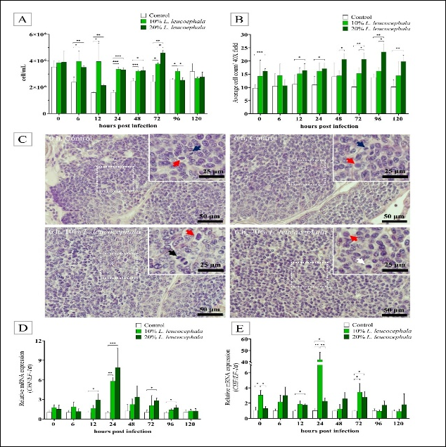
Figure 1. Hemocyte counts and hematopoietic cell proliferation in Macrobrachium rosenbergii fed fermented Leucaena leucocephala and infected with Aeromonas hydrophila. (A) Total hemocyte count at different time points post-infection (n = 5). (B) Average number of mitotic hematopoietic cells at different time points post-infection (n = 5). (C) Representative hematopoietic tissue at 6 h post-infection undergoing cell division; red arrow = prophase, white arrow = metaphase, black arrow = anaphase, and blue arrow = telophase. (D) Relative mRNA expression of CHF in hemocytes. (E) Relative mRNA expression of CHF in hematopoietic tissue. Data are expressed as mean ± SD. For gene expression analyses, n = 3 (cDNA pooled from two prawns per sample). *p < 0.05; **p < 0.01; ***p < 0.001.
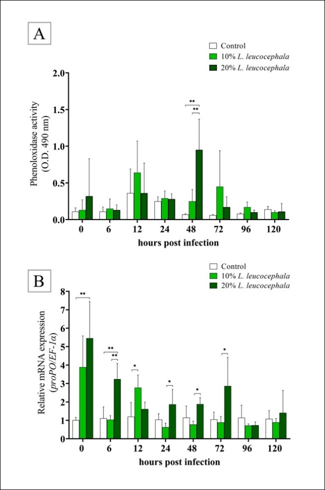
Figure 2. Effects of dietary fermented Leucaena leucocephala on phenoloxidase activity and prophenoloxidase expression in Macrobrachium rosenbergii infected with Aeromonas hydrophila. (A) Phenoloxidase activity in hemolymph. (B) Relative mRNA expression of prophenoloxidase in hemocytes. Data are expressed as mean ± SD. For gene expression analyses, n = 3 (cDNA pooled from two prawns per sample). *p < 0.05; **p < 0.01; ***p < 0.001.
Immune-related gene expression in hemocytes and hematopoietic tissue
The mRNA expression levels of TRAF6 and Dorsal in hemocytes were significantly lower in the 10% and 20% L. leucocephala groups than in the control group during 6–48 h post-infection (Figure 3A and B). At 72 h, TRAF6 expression remained downregulated in both treatment groups; however, the differences were no longer statistically significant compared with the control group. TRAF6 expression subsequently returned to levels comparable to those of the control group at 96–120 h post-infection (Figure 3A). Similarly, Dorsal mRNA expression was downregulated in the 10% and 20% L. leucocephala groups during 6–72 h and then returned to near control levels at 96–120 h post-infection (Figure 3B).
In hematopoietic tissue, TRAF6 expression did not differ significantly among the three groups throughout the experimental period (Figure 3C). In contrast, Dorsal expression in hematopoietic tissue was significantly decreased at 6 h in the 10% L. leucocephala group (0.21 ± 0.04) compared with the 20% L. leucocephala group (0.91 ± 0.19) and the control group (1.04 ± 0.34) (p < 0.01, Figure 3D). Although a general trend toward reduced Dorsal expression was observed at other time points, significant reductions were detected only in the 20% L. leucocephala group at 12 h (0.60 ± 0.08) and 72 h (0.64 ± 0.12), and in the 10% L. leucocephala group at 96 h (0.46 ± 0.10), compared with the control group (1.09 ± 0.49) (Figure 3D).
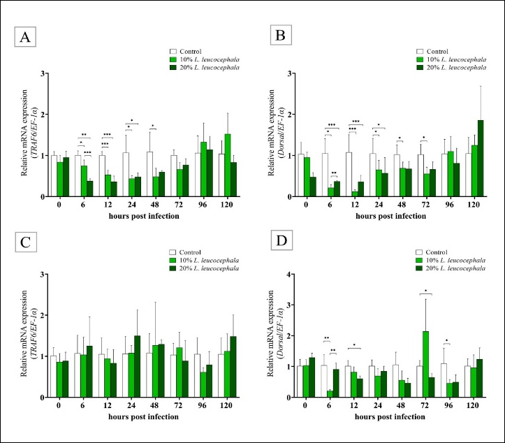
Figure 3. Expression of Toll/Dorsal pathway genes in hemocytes and hematopoietic tissue of Macrobrachium rosenbergii infected with Aeromonas hydrophila. (A) Relative mRNA expression of TRAF6 in hemocytes. (B) Relative mRNA expression of Dorsal in hemocytes. (C) Relative mRNA expression of TRAF6 in hematopoietic tissue. (D) Relative mRNA expression of Dorsal in hematopoietic tissue. Data are expressed as mean ± SD. For gene expression analyses, n = 3 (cDNA pooled from two prawns per sample). *p < 0.05; **p < 0.01; ***p < 0.001.
Overall, IMD expression was elevated in the 10% and 20% L. leucocephala groups, although expression patterns varied across time points (Figure 4A). At 0 h, IMD expression in hemocytes did not differ significantly between the 10% L. leucocephala group and the control group. However, the 20% L. leucocephala group (7.25 ± 1.74) showed significantly higher IMD expression than both the 10% L. leucocephala group (1.46 ± 0.29) (p < 0.05) and the control group (0.99 ± 0.6) (p < 0.01) (Figure 4A). During A. hydrophila infection, IMD expression remained upregulated in the 10% L. leucocephala group, although the increase was not statistically significant compared with the control group. In contrast, the 20% L. leucocephala group (2.20 ± 0.01) exhibited significantly higher IMD expression at 6 h post-infection than the control group (1.14 ± 0.63) (p < 0.05) (Figure 4A). IMD expression further increased in both treatment groups during 12–24 h post-infection, with the highest expression observed at 12 h (Figure 4A). Thereafter, although IMD expression remained higher than that in the control group, statistically significant differences were observed only at 72 h in both the 10% and 20% L. leucocephala groups (Figure 4A).
Relish expression was significantly increased in the 20% L. leucocephala group at 0 h (1.40 ± 0.08) compared with the control group (1.01 ± 0.17) (p < 0.05) (Figure 4B). At 6 h after bacterial challenge, Relish expression was significantly elevated in both the 10% (11.64 ± 5.49) and 20% L. leucocephala groups (12.69 ± 4.93) compared with the control group (1.13 ± 0.67) (p < 0.05) (Figure 4B). At 12 h, significantly higher Relish expression was observed only in the 20% L. leucocephala group (1.77 ± 0.10) compared with the 10% L. leucocephala group (1.23 ± 0.11) and the control group (1.04 ± 0.35) (p < 0.05) (Figure 4B). At 24 h, Relish expression was significantly upregulated in the 10% L. leucocephala group compared with the control group (p < 0.05) (Figure 4B). Although slight increases in Relish expression were observed in both treatment groups during 48–96 h post-infection, the differences were not statistically significant. At 120 h, Relish expression was significantly higher in the 20% L. leucocephala group (3.19 ± 1.04) than in the control group (1.00 ± 0.17) (p < 0.05) (Figure 4B).
HSP70 expression was significantly higher in the 20% L. leucocephala group than in the 10% L. leucocephala and control groups throughout the 0–120 h post-infection period (p < 0.001) (Figure 4C). HSP70 expression was highest at 0 h and gradually decreased until 48 h post-infection. Thereafter, expression gradually increased during 72–96 h, and by the end of the experimental period, no significant differences in HSP70 expression were observed among the groups.
Cu/Zn-SOD mRNA expression was significantly upregulated in the 20% L. leucocephala group compared with the 10% L. leucocephala and control groups during 0–12 h and at 120 h post-infection (p < 0.001) (Figure 4D). At 24 h post-infection, Cu/Zn-SOD expression was markedly elevated in both the 10% (50.44 ± 29.87) and 20% L. leucocephala groups (40.48 ± 26.46) compared with the control group (0.92 ± 0.17), although no significant difference was observed between the two treatment groups. During 72–120 h post-infection, both L. leucocephala-supplemented groups showed a trend toward increased Cu/Zn-SOD expression relative to the control group.
In addition, the marked upregulation of Cu/Zn-SOD expression in the 20% L. leucocephala group was associated with significantly lower ABTS and DPPH IC50 values than in the control group, indicating enhanced radical-scavenging activity. This group also exhibited higher FRAP values and increased TPC and TFC compared with the control group (Table 7). These findings suggest that elevated antioxidant gene expression corresponded with improved antioxidant capacity and phytochemical content.
| Diet | DPPH IC₅₀ ( μg /mL) | ABTS⁺ IC₅₀ ( μg /mL) | FRAP (mg FeSO ₄/g extract) | TPC (mg GAE/g extract) | TFC (mg CE/g extract) |
|---|---|---|---|---|---|
| 0% fermented L. leucocephala | 6,436.66 ± 88.19ᵃ | 1,604.48 ± 51.51ᵃ | 6.02 ± 0.50ᵃ | 2.40 ± 0.15ᵃ | −0.134 ± 0.1ᵃ |
| 10% fermented L. leucocephala | 2,189.23 ± 52.06ᵇ | 496.55 ± 12.64ᵇ | 15.13 ± 2.06ᵇ | 7.88 ± 0.42ᵇ | 0.882 ± 1.2ᵃ |
| 20% fermented L. leucocephala | 1,612.57 ± 19.87ᶜ | 305.38 ± 17.19ᶜ | 27.78 ± 2.23ᶜ | 11.62 ± 0.50ᶜ | 1.364 ± 0.4ᵃ |
Table 7. Antioxidant and phytochemical activities of experimental diets.
| Diet | DPPH IC₅₀ ( μg /mL) | ABTS⁺ IC₅₀ ( μg /mL) | FRAP (mg FeSO ₄/g extract) | TPC (mg GAE/g extract) | TFC (mg CE/g extract) |
|---|---|---|---|---|---|
| 0% fermented L. leucocephala | 6,436.66 ± 88.19ᵃ | 1,604.48 ± 51.51ᵃ | 6.02 ± 0.50ᵃ | 2.40 ± 0.15ᵃ | −0.134 ± 0.1ᵃ |
| 10% fermented L. leucocephala | 2,189.23 ± 52.06ᵇ | 496.55 ± 12.64ᵇ | 15.13 ± 2.06ᵇ | 7.88 ± 0.42ᵇ | 0.882 ± 1.2ᵃ |
| 20% fermented L. leucocephala | 1,612.57 ± 19.87ᶜ | 305.38 ± 17.19ᶜ | 27.78 ± 2.23ᶜ | 11.62 ± 0.50ᶜ | 1.364 ± 0.4ᵃ |
Values are presented as mean ± SD (n = 3). Different superscript letters within the same column indicate significant differences among treatments (p < 0.05). ABTS = 2,2′-azinobis-(3-ethylbenzothiazoline-6-sulfonic acid); CE = catechin equivalent; DPPH = 2,2-diphenyl-1-picrylhydrazyl; FRAP = ferric reducing antioxidant power; GAE = gallic acid equivalent; IC₅₀ = half-maximal inhibitory concentration; SD = standard deviation; TFC = total flavonoid content; TPC = total phenolic content; L. leucocephala = Leucaena leucocephala.
ALF mRNA expression was generally upregulated in the 10% and 20% L. leucocephala groups compared with the control group throughout the 0–120 h post-infection period (Figure 4E). The 20% L. leucocephala group exhibited significantly higher ALF expression than both the 10% L. leucocephala and control groups at 0 h (p < 0.01), 6 h (p < 0.01), 24 h (p < 0.05 and p < 0.01, respectively), and 120 h post-infection (p < 0.01) (Figure 4E). In contrast, ALF expression in the 10% L. leucocephala group (2.55 ± 0.32) was significantly higher than that in the control group (1.00 ± 0.12) only at 24 h post-infection (p < 0.05) (Figure 4E). In contrast to ALF, C-lectin expression was generally lower in the 10% and 20% L. leucocephala groups than in the control group at all time points. However, significant reductions were observed only during 6–12 h and 72–96 h post-infection (Figure 4F).
Intestinal morphology and intestinal immune-related gene expression
The histology of M. rosenbergii intestines showed normal structures in all groups (Figure 5A and Supplementary Figure S2). Alterations in epithelial cell height (Figure 5B), microvilli height (Figure 5C), and muscular wall thickness (Figure 5D) were not observed.
Overall, IMD expression was higher in the 20% L. leucocephala group than in the control and 10% L. leucocephala groups throughout the experimental period (Figure 6A). Compared with the control group, IMD expression in the 20% L. leucocephala group was significantly increased at 6 h (p < 0.05), 24 h (p < 0.001), and 48 and 96 h post-infection (p < 0.01), and at 120 h post-infection (p < 0.01). However, IMD expression in the 20% L. leucocephala group did not differ significantly from that in the 10% L. leucocephala group. In the 10% L. leucocephala group, IMD expression was significantly higher than that in the control group at 48 h (p < 0.05) and 120 h post-infection (p < 0.01) (Figure 6A).
Relish expression (Figure 6B) was generally higher in the 20% L. leucocephala group than in the control and 10% L. leucocephala groups throughout the experimental period. Specifically, Relish expression in the 20% L. leucocephala group was significantly elevated (p < 0.01) at 0 h (2.34 ± 0.34) compared with the control (1.04 ± 0.32) and 10% L. leucocephala groups (1.02 ± 0.35). Significant increases were also observed at 6 h (2.37 ± 0.77) compared with the control group (1.01 ± 0.17) and 10% L. leucocephala group (1.28 ± 0.21), at 48 h (2.29 ± 0.64) compared with both the control (1.01 ± 0.19) and 10% L. leucocephala groups (1.28 ± 0.21) (p < 0.01 and p < 0.05, respectively), and at 120 h (2.74 ± 0.50) compared with the control group (1.06 ± 0.43) and 10% L. leucocephala group (1.28 ± 0.62) (p < 0.01) post-infection. Although Relish expression in the 10% L. leucocephala group tended to be higher than that in the control group, significant differences were observed only at 24 h (2.25 ± 0.66) and 72 h (1.42 ± 0.25) post-infection (p < 0.05) (Figure 6B).
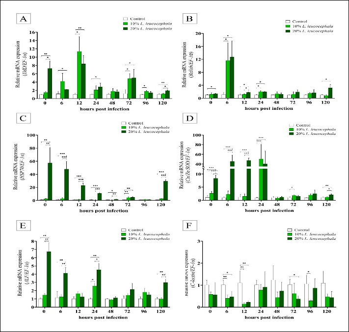
Figure 4. Effects of dietary fermented Leucaena leucocephala on IMD/Relish pathway gene expression in hemocytes of Macrobrachium rosenbergii infected with Aeromonas hydrophila. (A) Relative mRNA expression of IMD. (B) Relative mRNA expression of Relish. (C) Relative mRNA expression of HSP70. (D) Relative mRNA expression of Cu/Zn-SOD. (E) Relative mRNA expression of ALF. (F) Relative mRNA expression of C-lectin. Data are expressed as mean ± SD. For gene expression analyses, n = 4 (cDNA pooled from two prawns per sample). *p < 0.05; **p < 0.01; ***p < 0.001.
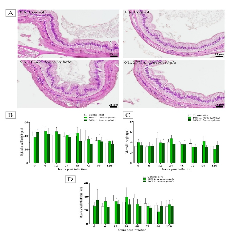
Figure 5. Effects of dietary fermented Leucaena leucocephala on intestinal morphology of Macrobrachium rosenbergii infected with Aeromonas hydrophila. (A) Representative hematoxylin and eosin-stained intestinal sections. (B) Epithelial cell height. (C) Microvillus height. (D) Muscular wall thickness. Data are expressed as mean ± SD (n = 5).
HSP70 expression (Figure 6C) was generally higher in the 20% L. leucocephala group than in the control and 10% L. leucocephala groups throughout the experimental period. At 0 h, HSP70 expression in the 20% L. leucocephala group (3.62 ± 0.84) was significantly higher than that in the control (1.03 ± 0.31) and 10% L. leucocephala groups (1.46 ± 0.67) (p < 0.001 and p < 0.01, respectively). At 6 h post-infection, HSP70 expression in the 20% L. leucocephala group (2.59 ± 0.98) was significantly higher than that in the control group (1.00 ± 0.14) (p < 0.05). Markedly elevated expression was also observed at 12 h (53.11 ± 20.19) and 24 h (16.77 ± 6.19) (p < 0.001), as well as at 96 h (7.33 ± 5.00) and 120 h (6.97 ± 1.03) post-infection compared with the control group (p < 0.001).
In the 10% L. leucocephala group, HSP70 expression tended to be higher than that in the control group, although significant differences were observed only at 6 h (4.36 ± 0.85) and 120 h (5.83 ± 1.73) post-infection (p < 0.001). Notably, at 6 h post-infection, HSP70 expression was higher in the 10% L. leucocephala group than in the 20% L. leucocephala group.
Cu/Zn-SOD expression (Figure 6D) was generally higher in the 20% L. leucocephala group than in the control and 10% L. leucocephala groups throughout the experimental period. Significant upregulation in the 20% L. leucocephala group was observed at 0 h (7.02 ± 2.93) compared with the control group (1.07 ± 0.49) (p < 0.001) and the 10% L. leucocephala group (3.04 ± 0.30) (p < 0.01). At 6 h post-infection, Cu/Zn-SOD expression increased further to 15.32 ± 2.38 and remained significantly higher than in the control group (p < 0.001). Significant increases were also observed at 24 h (3.25 ± 1.33) compared with both the control and 10% L. leucocephala groups (p < 0.01 and p < 0.05, respectively), and at 48 h (3.79 ± 2.28) compared with the control group (1.06 ± 0.40) (p < 0.05).
In the 10% L. leucocephala group, Cu/Zn-SOD expression also tended to be higher than that in the control group, although the increase was less pronounced than that observed in the 20% L. leucocephala group. Significant differences between the 10% L. leucocephala and control groups were detected only at 6, 24, and 96 h post-infection (p < 0.01).
ALF expression (Figure 6E) was generally higher in the 10% and 20% L. leucocephala groups than in the control group. Significant upregulation was observed at 0, 12, 48, 72, and 120 h post-infection in both treatment groups compared with the control group, although no significant differences were detected between the 10% and 20% L. leucocephala groups at these time points. At 6 h post-infection, ALF expression in the 20% L. leucocephala group (18.44 ± 16.15) also showed an increasing trend; however, the difference was not statistically significant compared with the control group. In contrast, the 10% L. leucocephala group (23.01 ± 4.31) exhibited significantly higher ALF expression than the control group (1.89 ± 1.86) at this time point.
C-lectin expression (Figure 6F) at 0 h was significantly lower in the 10% L. leucocephala (0.16 ± 0.07) and 20% L. leucocephala groups (0.24 ± 0.17) than in the control group (1.04 ± 0.33). However, at 12 h and 48 h post-infection, C-lectin expression was markedly elevated in the 20% L. leucocephala group, reaching 52.56 ± 10.57 and 11.24 ± 3.54, respectively, which were significantly higher than those in the control group (1.03 ± 0.26 and 1.04 ± 0.37, respectively) and the 10% L. leucocephala group (7.60 ± 2.58 and 1.81 ± 1.58, respectively) (p < 0.001).
C-lectin expression in the 10% L. leucocephala group also showed an increasing trend during the 12–96 h post-infection period, similar to that observed in the 20% L. leucocephala group. At 72 h, expression in the 10% L. leucocephala group (30.26 ± 6.47) was significantly higher than that in the control group (1.01 ± 0.20) and the 20% L. leucocephala group (6.27 ± 2.26) (p < 0.001). After 72 h post-infection, C-lectin expression in the 20% L. leucocephala group gradually decreased, resulting in a significant difference compared with the 10% L. leucocephala group (p < 0.05).
Mortality rates
The effect of 10% and 20% fermented L. leucocephala in fishmeal replacement on the survival of adult M. rosenbergii during A. hydrophila infection is shown in Figure 7. Survival of M. rosenbergii tended to be higher in the 10% and 20% L. leucocephala groups than in the control group; however, the differences were not statistically significant (p = 0.167 and p = 0.204, respectively). No dose-dependent effect was observed.
DISCUSSION
Functional value of fermented L. leucocephala as a fishmeal substitute
L. leucocephala leaves are widely recognized as a rich source of protein and essential minerals, making them a promising alternative feed ingredient for livestock and aquaculture species in tropical regions. Their high protein content is a key nutritional advantage, along with the presence of 17 flavonoids known for biological activities, including antioxidant and anti-inflammatory effects [14]. In the present study, L. plantarum TISTR1284 was used to improve the bioavailability and functional properties of L. leucocephala leaves. Fermentation is known to enhance food safety and nutritional quality by producing organic acids, including lactic, acetic, propionic, and formic acids, which help eliminate harmful bacteria and toxins while preserving sensory qualities [17]. However, the present experimental design does not allow separation of the individual contributions of fermentation, plant substitution, and probiotic effects; therefore, the outcomes observed here should be interpreted as the combined effect of the fermented ingredient rather than being attributed to a single factor.
A. hydrophila, a pathogenic Gram-negative bacterium, poses a serious threat to Macrobrachium nipponense and M. rosenbergii [34, 35]. Plant extracts from Withania somnifera and Melaleuca cajuputi have been shown to enhance immune responses and increase shrimp survival during A. hydrophila infection [36, 37]. L. leucocephala seed protein has also been shown to enhance bacterial disease resistance in the catfish Clarias gariepinus [12]. In the present study, dietary supplementation with fermented L. leucocephala leaves enhanced immune parameters and improved survival of M. rosenbergii following A. hydrophila challenge.
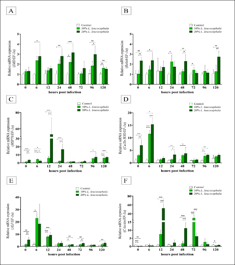
Figure 6. Effects of dietary fermented Leucaena leucocephala on IMD/Relish pathway and immune-related gene expression in the intestine of Macrobrachium rosenbergii infected with Aeromonas hydrophila. (A) Relative mRNA expression of IMD. (B) Relative mRNA expression of Relish. (C) Relative mRNA expression of HSP70. (D) Relative mRNA expression of Cu/Zn-SOD. (E) Relative mRNA expression of ALF. (F) Relative mRNA expression of C-lectin. Data are expressed as mean ± SD. For gene expression analyses, n = 4 (cDNA pooled from two prawns per sample). *p < 0.05; **p < 0.01; ***p < 0.001.
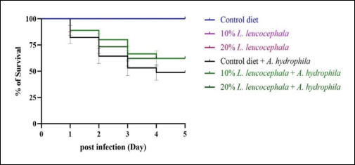
Figure 7. Survival rate of Macrobrachium rosenbergii following Aeromonas hydrophila infection. Data are expressed as mean ± SD (n = 3 replicate tanks per group). Survival analysis was performed using the log-rank (Mantel–Cox) test. No significant differences were observed between the control and 10% replacement groups (p = 0.167) or between the control and 20% replacement groups (p = 0.204).
Hemocyte proliferation and hematopoietic activity
Crustacean resistance to pathogens relies primarily on the innate immune system. Hemocytes are crucial cells that mediate physiological responses to infection and environmental stress. Probiotics and medicinal plant extracts have been applied to enhance immune parameters in M. rosenbergii [38, 39]. In the present study, increased circulating hemocytes and a higher number of mitotic cells in hematopoietic tissue were observed when fermented L. leucocephala leaves were added to the diet of M. rosenbergii. This response may contribute to accelerated maturation of hemocyte precursors in hematopoietic tissue, thereby maintaining hemocyte population and functionality during bacterial challenge [40].
L. leucocephala contains apigenin, kaempferol, and juglanin, which induce the transcriptional activity of pHRE-Luc before promoting erythropoietin expression and stimulating red blood cell production [14]. Upregulation of CHF is a biological indicator associated with hemocyte and hematopoietic cell proliferation in M. rosenbergii [27]. The increase in hematopoietic proliferation, reflected by CHF expression in hemocytes and hematopoietic tissue of the 10% and 20% L. leucocephala groups, suggests that fermented L. leucocephala promotes hematopoietic activity and hemocyte homeostasis during bacterial infection.
PO activity and proPO expression
PO is an important enzyme that promotes humoral defense mechanisms and the elimination of pathogens in invertebrates [41]. Plant extracts have been shown to induce PO activity and improve immunocompetence against pathogens in crustaceans [38, 42, 43]. In the present study, M. rosenbergii fed fermented L. leucocephala showed increased PO activity in hemolymph and significant upregulation of proPO. Antimicrobial and antioxidative activities induced by phytochemicals in L. leucocephala have been previously reported [44]. These findings suggest that key bioactive properties of L. leucocephala were retained after fermentation, although the specific compounds responsible for the observed effects were not directly quantified in the present study.
Modulation of Toll pathway-related signaling
TRAF6 is a highly expressed gene that acts as a crucial signal transducer triggering downstream cascades mediated by TNF receptors and the IL-1 receptor/Toll-like receptor superfamily during immune responses [45]. In Fenneropenaeus penicillatus, higher TRAF6 expression was correlated with peroxinectin expression after infection by white spot syndrome virus and Vibrio alginolyticus [46]. TRAF6-like transcription was upregulated after Staphylococcus aureus and Edwardsiella ictaluri challenge in Procambarus clarkii [47]. Knockdown of TRAF6-like inhibited downstream effector genes, including Dorsal [47]. TRAF6 regulates immune responses during A. hydrophila infection because mannose-binding lectin and crustin are decreased after TRAF6 silencing [30].
In the present study, TRAF6 expression decreased within 48 h, and Dorsal expression declined at specific time points in the 10% and 20% groups. These findings indicate modulation of inflammatory signaling; however, bacterial burden or clearance was not quantified in this study. Therefore, improved survival should not be directly interpreted as increased bacterial clearance. Flavonoids in L. leucocephala have been reported to reduce pro-inflammatory cytokine secretion in RAW 264.7 macrophages [14], which may partly explain the moderated inflammatory profile observed here.
IMD pathway and immune-related gene responses
Two major pathways are involved in shrimp immunity: the Toll pathway and the IMD pathway. These pathways share downstream signaling components, and crosstalk between them contributes to protection against bacterial infection [48]. In the present study, C-lectin was downregulated, whereas IMD-related genes (IMD and Relish) and effector/stress genes (HSP70, Cu/Zn-SOD, and ALF) were upregulated, particularly in the 20% group. This pattern suggests coordinated immune modulation rather than simple immune suppression. Such modulation may reflect regulated immune adjustment to dietary bioactive compounds, as previously observed in crustaceans exposed to functional feed additives and plant-derived extracts [49, 50].
However, the precise molecular mechanism underlying the activation of the immune pathway by fermented L. leucocephala remains unclear and warrants further mechanistic investigation. In particular, this study did not identify the specific bioactive compounds responsible for modulating the IMD pathway, although phytochemicals such as phenolics have been reported to influence immune responses in C. gariepinus [51]. Nevertheless, the exact molecular mediators and their direct interactions with IMD pathway components remain unclear and require targeted metabolomic and functional studies.
Gut health, paraprobiotic effects, and postbiotic contributions
In addition to immunomodulation, L. leucocephala has been associated with gut health and nutrient utilization [52]. Replacing 25%–50% of fishmeal with L. leucocephala leaves in Nile tilapia diets did not adversely affect fish health, although some growth retardation was observed [11]. Improvements in digestibility and nutrient uptake have also been reported when L. leucocephala leaves are fermented with beneficial gut bacteria. For example, Bacillus subtilis and Bacillus circulans enhanced nutritional value and growth performance in Labeo rohita when included in diets containing 30%–40% L. leucocephala [53]. In crustaceans, host-associated probiotics such as Lactococcus lactis have shown similar benefits in M. rosenbergii, improving both growth and immune function [39].
Although viable LAB counts in the final pelleted feed and during storage were not quantified, the present findings suggest that the beneficial effects observed in M. rosenbergii were likely mediated not only by live probiotic cells but also by fermentation-derived paraprobiotics and postbiotics acting synergistically with enhanced plant bioactive compounds. Paraprobiotics, including inactivated microbial cells and cell-wall components such as peptidoglycans, teichoic acids, and surface proteins, are known to interact with crustacean pattern-recognition receptors, thereby stimulating hemocyte proliferation and activating downstream immune pathways, including the IMD–Relish signaling cascade observed in the present study. Concurrently, fermentation by L. plantarum may generate beneficial postbiotic metabolites, including organic acids, bacteriocins, and bioactive intracellular compounds, which may further contribute to immune modulation and disease resistance.
Detoxification and antioxidant-related benefits
The present findings suggest that L. plantarum fermentation may help mitigate one of the major limitations associated with plant protein utilization, namely ANFs. As shown in Table 1, fermentation reduced mimosine levels by 42.18%, decreasing from 92.46 to 53.46 mg/100 g. Mimosine is a toxic non-protein amino acid known to impair nutrient utilization and physiological performance in animals. Furthermore, the incorporation of fermented L. leucocephala enhanced the phytochemical and antioxidant properties of the diets, as evidenced by increased TPC and TFC, higher FRAP values, and improved radical-scavenging activity, as indicated by lower DPPH and ABTS IC50 values in the 20% inclusion group. Collectively, these findings suggest that fermented diets may function as promising functional feeds enriched with detoxified plant matrixes, fermentation-derived metabolites, and microbial-derived bioactive components that collectively support innate immune responses in prawns.
Study limitations and future research
One limitation of the present study is that the experimental diets were not strictly formulated to be isonitrogenous. As shown in Table 2, crude protein content decreased from 44.63% in the control diet to 36.75% in the 20% replacement group, representing a substantial nutritional variation that may have influenced the physiological and immunological responses of M. rosenbergii. Therefore, the effects cannot be attributed exclusively to the bioactive properties of fermented L. leucocephala, as differences in dietary protein levels may also have contributed to the observed responses. In addition, crystalline amino acids such as lysine and methionine were not supplemented to achieve precise amino acid balancing among the experimental diets. Consequently, variations in essential amino acid composition resulting from the inclusion of fermented L. leucocephala may have further influenced immune and physiological responses. Future studies using strictly isonitrogenous, isolipidic, and amino acid-balanced diets are required to better distinguish the specific functional effects of fermented L. leucocephala from potential nutritional confounding factors.
Another limitation is that the feeding trial duration was relatively short compared with conventional aquaculture nutrition studies, and growth performance parameters such as weight gain, specific growth rate, and feed conversion ratio were not evaluated. In addition, pellet water stability and nutrient leaching rates were not quantitatively assessed. Since feed stability in water may influence nutrient availability, feed intake, and feeding efficiency in prawns, these parameters should be evaluated in future studies to better characterize the physicochemical quality and practical applicability of the experimental diets under aquaculture conditions. Therefore, the long-term nutritional suitability and commercial applicability of the diets could not be fully determined in the present study. Furthermore, the current experimental design does not allow precise determination of the optimal inclusion level or dose-dependent immune responses associated with fermented L. leucocephala supplementation.
The present study also has limitations related to fermentation characterization and microbial analysis. Post-fermentation LAB viability, pH, lactic acid concentration, titratable acidity, and detailed biochemical characteristics of the fermented product were not directly quantified. In addition, the microbial composition of the fermented material and viability of L. plantarum in the final feed and during storage were not evaluated. Consequently, microbial dynamics and fermentation-derived metabolites associated with the fermented product could not be fully characterized. Therefore, the relative contributions of live probiotic cells, postbiotic metabolites, and paraprobiotic effects remain unclear. Moreover, microbial safety analyses, including assessments of pathogen and mycotoxin contamination, were not performed directly, although fermentation was conducted under sealed, anaerobic conditions to minimize contamination risk. Future studies integrating comprehensive fermentation profiling, microbial safety analyses, metabolite characterization, and gut microbiota analysis would provide stronger mechanistic insight into host–microbe interactions and the functional properties of fermented L. leucocephala.
Antioxidant capacity was not directly measured in hemolymph or tissues of M. rosenbergii. The present study primarily evaluated antioxidative responses through immune- and oxidative stress-related gene expression, particularly Cu/Zn-SOD expression in hemocytes. Therefore, the antioxidative effects of fermented L. leucocephala supplementation should be interpreted as indirect molecular evidence rather than direct biochemical confirmation of antioxidant activity.
Several limitations were also associated with the bacterial challenge experiment. Unchallenged control prawns were not sampled at all post-challenge time points (6–120 h), limiting the ability to distinguish the direct effects of dietary supplementation from diet × infection interaction effects. In addition, bacterial load in hemolymph or tissues was not quantified. Therefore, the improved survival and immune responses observed in the treatment groups cannot be directly interpreted as evidence of enhanced bacterial clearance capacity. Correlation analyses between immune parameters and survival outcomes were also not performed, preventing detailed evaluation of the quantitative relationship between immune modulation and disease resistance. Future studies incorporating time-matched unchallenged controls, quantification of bacterial load, and integrated multivariate statistical analyses would strengthen the mechanistic interpretation of immune responses and disease resistance in M. rosenbergii.
Despite these limitations, the consistent enhancement of several immune parameters and improved resistance to A. hydrophila observed in the treatment groups suggest that fermented L. leucocephala may contribute beneficial immunomodulatory effects in prawns. Importantly, no adverse effects on intestinal morphology were detected, supporting the safety of the dietary inclusion levels used in the present study. Nevertheless, the findings should be interpreted with caution, and further long-term studies using rigorously controlled diet formulations and comprehensive physiological, microbiological, and biochemical evaluations are warranted to clarify the specific contribution of fermented L. leucocephala to immune modulation and disease resistance in prawn aquaculture.
CONCLUSION
Dietary supplementation with fermented L. leucocephala enhanced several innate immune responses in M. rosenbergii challenged with A. hydrophila. Fermentation improved the nutritional quality of L. leucocephala by increasing essential amino acid content and reducing mimosine concentration, while supplementation of experimental diets increased TPC, TFC, antioxidant activity, and reducing power. Prawns receiving fermented L. leucocephala, particularly at the 20% inclusion level, exhibited enhanced hemocyte proliferation, increased hematopoietic activity, elevated PO activity, and upregulated expression of key immune- and stress-related genes, including CHF, proPO, IMD, Relish, HSP70, Cu/Zn-SOD, and ALF. These responses were accompanied by improved resistance to A. hydrophila infection and the maintenance of normal intestinal morphology, indicating that the evaluated dietary inclusion levels were well tolerated.
From a practical perspective, the findings suggest that fermented L. leucocephala has potential as a functional feed ingredient and partial fishmeal substitute for freshwater prawn culture, providing both nutritional and immunological benefits. The study further demonstrates that fermentation with L. plantarum may reduce ANFs while enhancing the bioactive and antioxidant properties of plant-derived feed ingredients.
A major strength of this study is the comprehensive evaluation of immune responses through hematological, histological, molecular, and antioxidant-related assessments following bacterial challenge.
Future studies should employ isonitrogenous and amino acid-balanced diets, evaluate long-term growth and production performance, characterize fermentation-derived metabolites and microbial dynamics, quantify pathogen clearance, and investigate gut microbiota responses. Such studies will help clarify the mechanisms underlying immune modulation and establish the optimal inclusion level of fermented L. leucocephala for commercial aquaculture applications.
Overall, the present study provides evidence that fermented L. leucocephala can serve as a promising functional feed ingredient that enhances innate immunity, antioxidant status, and disease resistance in M. rosenbergii, supporting its potential application in sustainable prawn aquaculture.
DATA AVAILABILITY
The supplementary data can be made available from the corresponding author upon request.
GENERATIVE AI DECLARATION
The authors declare that generative artificial intelligence (AI) tools were used solely to improve language, grammar, and readability during manuscript preparation. All scientific content, data analysis, interpretation of results, and conclusions were developed and verified by the authors. The authors take full responsibility for the accuracy, integrity, and originality of the work presented, and no AI tool was listed as an author.
AUTHORS’ CONTRIBUTIONS
AP: Methodology, investigation, validation, formal analysis, data curation, visualization, and manuscript drafting. LP and KJ: Methodology, investigation, and data curation. SlS, SkS, PaP, and PoP: Bacterial culture and feed pellet preparation. SuS: Formal analysis and data curation. RV and CC: Validation, formal analysis, visualization, conceptualization, manuscript review, and editing. All authors have read and approved the final manuscript.
COMPETING INTERESTS
The authors declare that they have no competing interests.
PUBLISHER’S NOTE
Veterinary World remains neutral with regard to jurisdictional claims in the published institutional affiliations.
ACKNOWLEDGMENTS
The authors gratefully acknowledge financial support from the Fundamental Fund under the Thailand Science Research and Innovation (TSRI) program and the University of Phayao (Grant No. FF64-UoE24). Additional support was provided by the National Science, Research and Innovation Fund (NSRF) through the Program Management Unit for Human Resources & Institutional Development, Research and Innovation (Grant No. B05F640137). Sukanya Saedan received a Scholarship for the Development of High-Quality Research Graduates in Science and Technology jointly administered by the National Science and Technology Development Agency (NSTDA) and Mahidol University.
The authors also sincerely thank Asst. Prof. Amnart Onsa-Ard and Miss Piyawan Nuntaboon, Division of Biochemistry, School of Medical Sciences, University of Phayao, Thailand, for their invaluable technical assistance with antioxidant and phytochemical assays.
REFERENCES
- Xiao F, Wang J, Liu H, Zhuang M, Wen X, Zhao H, Wu K. Effects of dietary protein levels on growth, digestive enzyme activity, antioxidant capacity, and gene expression related to muscle growth and protein synthesis of juvenile greasyback shrimp (Metapenaeus ensis). Animals 2023;13(24):3886. [Google Scholar]
- D'Abramo LR, Sheen SS. Nutritional requirements, feed formulation, and feeding practices for intensive culture of the freshwater prawn Macrobrachium rosenbergii. Rev Fish Sci 1994;2(1):1-21. [Google Scholar]
- Daniel N. A review on replacing fish meal in aqua feeds using plant protein sources. Int J Fish Aquat Stud 2018;6(2):164-179. [Google Scholar]
- Wang H, Hu X, Chen J, Zheng Y, Tan B, Shi L, Zhang S. Impact of dietary protein levels on growth, immune response, digestibility, intestinal microbiota, and transcriptome in Litopenaeus vannamei fed cottonseed protein concentrate. Aquac Rep 2025;42:102762. [Google Scholar]
- Macias-Sancho J, Poersch LH, Bauer W, Romano LA, Wasielesky W, Tesser MB. Fishmeal substitution with Arthrospira (Spirulina platensis) in a practical diet for Litopenaeus vannamei: effects on growth and immunological parameters. Aquaculture 2014;426:120-125. [Google Scholar]
- Hua K, Cobcroft JM, Cole A, Condon K, Jerry DR, Mangott A, Praeger C, Vucko MJ, Zeng C, Zenger K, Strugnell JM. The future of aquatic protein: implications for protein sources in aquaculture diets. One Earth 2019;1(3):316-329. [Google Scholar]
- Naylor RL, Hardy RW, Bureau DP, Chiu A, Elliott M, Farrell AP, Forster I, Gatlin DM, Goldburg RJ, Nichols PD. Feeding aquaculture in an era of finite resources. PNAS 2009;106(36):15103-15110. [Google Scholar]
- Hardy RW. Utilization of plant proteins in fish diets: effects of global demand and supplies of fishmeal. Aquac Res 2010;41(5):770-776. [Google Scholar]
- Lan TTP, To TTH. Effects of replacing fish meal with different levels of Lead tree (Leucaena leucocephala) leaf powder on growth, survival, digestive enzymes activity, muscle biochemical composition and texture of white-leg shrimp Penaeus vannamei Boone, 1931. Indian J Fish 2023;70(1):57-63. [Google Scholar]
- Ekpenyong TE. Nutrient and amino acid composition of Leucaena leucocephala (Lam.) de Wit. Anim Feed Sci Technol 1983;15(3):183-187. [Google Scholar]
- Wee KL, Wang SS. Nutritive value of Leucaena leaf meal in pelleted feed for Nile tilapia. Aquaculture 1987;62(2):97-108. [Google Scholar]
- Verma VK, Rani KV, Kumar SR, Prakash O. Leucaena leucocephala pod seed protein as an alternate to animal protein in fish feed and evaluation of its role to fight against infection caused by Vibrio harveyi and Pseudomonas aeruginosa. Fish Shellfish Immunol 2018;76:324-332. [Google Scholar]
- Haggag EG, Kamal AM, Abdelhady MIS, El-Sayed MM, El-Wakil EA, Abd-El-hamed SS. Antioxidant and cytotoxic activity of polyphenolic compounds isolated from the leaves of Leucaena leucocephala. Pharm Biol 2011;49:1103-1113. [Google Scholar]
- Xu Y, Tao Z, Jin Y, Yuan Y, Dong TT, Tsim KW, Zhou Z. Flavonoids, a potential new insight of Leucaena leucocephala foliage in ruminant health. J Agric Food Chem 2018;66(29):7616-7626. [Google Scholar]
- Wachid BAA, Setyowati DNA, Azhar F. Effectiveness of meniran leaf extract (Phyllanthus niruri L.) as immunostimulant in Vannamei Shrimp (Litopenaeus vannamei) against vibriosis disease. JAFH 2022;11(2):182-192. [Google Scholar]
- Serra V, Pastorelli G, Tedesco DEA, Turin L, Guerrini A. Alternative protein sources in aquafeed: Current scenario and future perspectives. 2024. [Google Scholar]
- Aleman RS, Montero-Fernández I, Marcía JA, Saravia Maldonado SA, Martín-Vertedor D. Application of fermentation as a strategy for the transformation and valorization of vegetable matrixes. Fermentation 2024;10(3):124. [Google Scholar]
- Myo H, Nantarat N, Khat-Udomkiri N. Changes in bioactive compounds of coffee pulp through fermentation-based biotransformation using Lactobacillus plantarum TISTR 543 and its antioxidant activities. Fermentation 2021;7(4):292. [Google Scholar]
- Dash G, Raman RP, Prasad KP, Makesh M, Pradeep MA, Sen S. Evaluation of Lactobacillus plantarum as feed supplement on host associated microflora, growth, feed efficiency, carcass biochemical composition and immune response of giant freshwater prawn, Macrobrachium rosenbergii (de Man, 1879). Aquaculture 2014;432:225-236. [Google Scholar]
- Pudgerd A, Saedan S, Kreungkum T, Sritunyalucksana K, Sanpa S, Somnet S, Vanichviriyakit R, Chotwiwatthanakun C. Inducing bursicon expression using 20-hydroxyecdysone (20E) increased immune response in Macrobrachium rosenbergii against Aeromonas hydrophila. Biol Open 2025;14(7):bio061773. [Google Scholar]
- Ren H, Feng Y, Pei J, Li J, Wang Z, Fu S, Zheng Y, Li Z, Peng Z. Effects of Lactobacillus plantarum additive and temperature on the ensiling quality and microbial community dynamics of cauliflower leaf silages. Bioresour Technol 2020;307:123238. [Google Scholar]
- AOAC International. Official methods of analysis of AOAC International Rockville: AOAC International; 2019. [Google Scholar]
- Thim-uam A, Thepmalee C, Chaiwangyen W, Kangwan N, Chokchaisiri R, Nuntaboon P, Songkrao A, Onsa-ard A. Phytochemical screening and in vitro antioxidant and anticancer evaluation of stem and leaf extracts of Cissampelos pareira L. Mediators Inflamm 2025;2025(1):7555073. [Google Scholar]
- Chaiwangyen W, Khantamat O, Pintha K, Kangwan N, Onsa-ard A, Nuntaboon P, Songkrao A, Thippraphan P, Chaiyasit D, de Sousa FLP. Cleistocalyx nervosum var. paniala mitigates oxidative stress and inflammation induced by PM10 soluble extract in trophoblast cells via miR-146a-5p. Sci Rep 2024;14(1):24265. [Google Scholar]
- Ghosh AK, Bir J, Azad MAK, Hasanuzzaman AFM, Islam MS, Huq KA. Impact of commercial probiotics application on growth and production of giant fresh water prawn (Macrobrachium rosenbergii De Man, 1879). Aquac Rep 2016;4:112-117. [Google Scholar]
- Ramasamy SM, Denis M, Sivakumar S, Munusamy A. Phenoloxidase activity in humoral plasma, hemocyanin and hemocyanin separated proteins of the giant freshwater prawn Macrobrachium rosenbergii. Int J Biol Macromol 2017;102:977-985. [Google Scholar]
- Jariyapong P, Pudgerd A, Cheloh N, Hirono I, Kondo H, Vanichviriyakit R, Weerachatyanukul W, Chotwiwatthanakun C. Hematopoietic tissue of Macrobrachium rosenbergii plays dual roles as a source of hemocyte hematopoiesis and as a defensive mechanism against Macrobrachium rosenbergii nodavirus infection. Fish Shellfish Immunol 2019;86:756-763. [Google Scholar]
- Pudgerd A, Kruangkum T, Sritunyalucksana K, Vanichviriyakit R, Imsonpang S, Chotwiwatthanakun C. Immunopathogenesis of hematopoietic tissues in response to Vibrio parahaemolyticus (VPAHPND) infection in Macrobrachium rosenbergii. Fish Shellfish Immunol 2021;110:10-22. [Google Scholar]
- Sun C, Liu B, Zhou Q, Xiong Z, Shan F, Zhang H. Response of Macrobrachium rosenbergii to vegetable oils replacing dietary fish oil: Insights from antioxidant defense. Front Physiol 2020;11:218. [Google Scholar]
- Choolert C, Pasookhush P, Vaniksampanna A, Longyant S, Chaivisuthangkura P. A novel tumor necrosis factor receptor-associated factor 6 (TRAF6) gene from Macrobrachium rosenbergii involved in antibacterial defense against Aeromonas hydrophila. Fish Shellfish Immunol 2023;140:108945. [Google Scholar]
- Jariyapong P, Vanichviriyakit R, Chotwiwatthanakun C, Pudgerd A. Alteration of the haemocyte parameters, expression of immune‐related and neurohormone bursicon genes of giant river prawn Macrobrachium rosenbergii (de Man) in the response to salinity stress and ammonia stress. Aquac Res 2020;51(6):2573-2581. [Google Scholar]
- Zhao C, Wen H, Huang S, Weng S, He J. A novel disease (water bubble disease) of the giant freshwater prawn Macrobrachium rosenbergii caused by Citrobacter freundii: antibiotic treatment and effects on the antioxidant enzyme activity and immune responses. Antioxidants 2022;11(8):1491. [Google Scholar]
- Livak KJ, Schmittgen TD. Analysis of relative gene expression data using real-time quantitative PCR and the 2(-Delta Delta C(T)) method. Methods 2001;25(4):402-408. [Google Scholar]
- Chen Q, Zhang Z, Tang H, Zhou L, Ao S, Zhou Y, Zhu X, Gao X, Jiang Q, Tu C, Zhang X. Aeromonas hydrophila associated with red spot disease in Macrobrachium nipponense and host immune-related gene expression profiles. J Invertebr Pathol 2021;182:107584. [Google Scholar]
- Chu P, Qian Q, Shen Y, Zhu Y, Wang Y, Yao X, Zhuang M, Zhu L, Zhang X. Autophagy increases the survival rate of Macrobrachium rosenbergii after Aeromonas hydrophila infection. Aquaculture 2023;575:739758. [Google Scholar]
- Harikrishnan R, Balasundaram C, Jawahar S, Heo MS. Immunomodulatory effect of Withania somnifera supplementation diet in the giant freshwater prawn Macrobrachium rosenbergii (de Man) against Aeromonas hydrophila. Fish Shellfish Immunol 2012;32(1):94-100. [Google Scholar]
- Mohamad Khairul Sahimi MB, Nagi AM, Hamdan NA, Zakariah MI, Yusoff NAH, Victor Tosin O, Hassan M. Cajeput Melaleuca cajuputi extract supplementation in diets of Macrobrachium rosenbergii: Insight on the growth, immunological responses and resistance against Aeromonas hydrophila. Aquac Res 2022;53(9):3441-3452. [Google Scholar]
- Halim AM, Lee PP, Chang ZW, Chang CC. The hot-water extract of leaves of noni, Morinda citrifolia, promotes the immunocompetence of giant freshwater prawn, Macrobrachium rosenbergii. Fish Shellfish Immunol 2017;64:457-468. [Google Scholar]
- Kader MA, Zahidah Azahar N, Iehata S, Bulbul M, Islam MM, Sarker J, Rahman MM, Asaduzzaman M. Dietary supplementation of host‐associated lactic acid bacteria modulates growth, metabolic activities, and immune‐related gene expression in giant freshwater prawn, Macrobrachium rosenbergii. J World Aquac Soc 2021;52(1):216-230. [Google Scholar]
- Sequeira T, Tavares D, Arala-Chaves M. Evidence for circulating hemocyte proliferation in the shrimp Penaeus japonicus. Dev Comp Immunol 1996;20(2):97-104. [Google Scholar]
- Amparyup P, Charoensapsri W, Tassanakajon A. Prophenoloxidase system and its role in shrimp immune responses against major pathogens. Fish Shellfish Immunol 2013;34(4):990-1001. [Google Scholar]
- Lawhavinit OA, Sincharoenpoka P, Sunthornandh P. Effects of ethanol tumeric (Curcuma longa Linn.) extract against shrimp pathogenic Vibrio spp. and on growth performance and immune status of white shrimp (Litopenaeus vannamei). J Agric Nat Resour 2011;45(1):70-77. [Google Scholar]
- Jayanthi R, Malar HV, Charles PM. Effect of Phyllanthus niruri on Penaeus indicus post larvae against WSSV infection. Int J Fish Aquac 2013;3:130-135. [Google Scholar]
- Elbanoby NE, El-Settawy AA, Mohamed AA, Salem MZ. Phytochemicals derived from Leucaena leucocephala (Lam.) de Wit (Fabaceae) biomass and their antimicrobial and antioxidant activities: HPLC analysis of extracts. Biomass Convers Biorefin 2024;14(13):14593-14609. [Google Scholar]
- Ye H, Arron JR, Lamothe B, Cirilli M, Kobayashi T, Shevde NK, Segal D, Dzivenu OK, Vologodskaia M, Yim M, Du K. Distinct molecular mechanism for initiating TRAF6 signalling. Nature 2002;418(6896):443-447. [Google Scholar]
- Cai S, Huang Y, Wang B, Jian J, Xu Y. Tumor necrosis factor receptor-associated factor 6 (TRAF6) participates in peroxinectin gene expression in Fenneropenaeus penicillatus. Fish Shellfish Immunol 2017;64:193-201. [Google Scholar]
- Li B, Yang BB, Shen XL, Wang K, Wei Z, Du ZQ. Molecular characterization and expression analysis of tumor necrosis factor receptor-associated factor 6 (traf6) like gene involved in antibacterial innate immune of freshwater crayfish, Procambarus clarkii. Fish Shellfish Immunol 2020;104:517-526. [Google Scholar]
- Liu Y, Song L, Sun Y, Liu T, Hou F, Liu X. Comparison of immune response in Pacific white shrimp, Litopenaeus vannamei, after knock down of Toll and IMD gene in vivo. Dev Comp Immunol 2016;60:41-52. [Google Scholar]
- Lai GL, Lin YR, Yang TJ, Chu YT, Nan FH, Hu YF. Effects of feed supplementation with fermented Broussonetia papyrifera leaves on non-specific immune responses, resistance to Vibrio parahaemolyticus, growth, intestinal histology, and microbiota of white shrimp (Penaeus vannamei). Fish Shellfish Immunol 2025;167:110901. [Google Scholar]
- Kuo YC, Tran HTQ, Ho TH, Bharadwaj A, Chu YT, Chen TY, Nan FH, Lee PT. Effects of dietary corn-fermented protein as a sustainable ingredient on growth performance, immune response, and disease resilience in whiteleg shrimp (Litopenaeus vannamei). Fish Shellfish Immunol 2026;174:111340. [Google Scholar]
- Verma VK, Rani KV, Sehgal N, Prakash O. Immunostimulatory effect of artificial feed supplemented with indigenous plants on Clarias gariepinus against Aeromonas hydrophila. Fish Shellfish Immunol 2013;35(6):1924-1931. [Google Scholar]
- Guo X, Qian Z, Pan Q, Hu Y, Mei W, Xing X, Yin S, Ji J, Zhang K. Effects of florfenicol on intestinal histology, apoptosis and gut microbiota of Chinese mitten crab (Eriocheir sinensis). Int J Mol Sci 2023;24(5):4412. [Google Scholar]
- Bairagi A, Sarkar Ghosh K, Sen SK, Ray AK. Evaluation of the nutritive value of Leucaena leucocephala leaf meal, inoculated with fish intestinal bacteria Bacillus subtilis and Bacillus circulans in formulated diets for rohu, Labeo rohita (Hamilton) fingerlings. Aquac Res 2004;35(5):436-446. [Google Scholar]