
ABSTRACT
Background and Aim: Food-producing animals are recognized reservoirs of antimicrobial-resistant bacteria with zoonotic potential. Third-generation cephalosporin-resistant (3GC-R) Escherichia coli is of particular public health concern due to its association with extended-spectrum β-lactamase (ESBL) and plasmid-mediated AmpC enzymes. This study aimed to determine the prevalence, resistance mechanisms, transferability of resistance genes, efflux pump contribution, and genetic relatedness of 3GC-R E. coli isolated from farm animals and workers on a small-scale farm in lower northern Thailand.
Materials and Methods: A total of 265 fecal samples were collected from laying hens (n = 210), cattle (n = 33), swine (n = 19), and farm workers (n = 3). Isolation of 3GC-R E. coli was performed using cefotaxime-supplemented selective media. Antimicrobial susceptibility was determined by disk diffusion and minimum inhibitory concentration (MIC) assays. The presence of blaCTX-M and blaCMY-2 genes was identified by polymerase chain reaction and sequencing. Conjugation assays and plasmid replicon typing assessed gene transferability. Efflux pump involvement in ceftazidime resistance was evaluated using phenylalanine-arginine β-naphthylamide. Genetic relatedness was analyzed by pulsed-field gel electrophoresis.
Results: Overall, 15.8% of samples yielded 3GC-R E. coli, with the highest prevalence observed in swine (47.4%), followed by cattle (27.3%) and laying hens (10.5%). Multidrug resistance was detected in 57.1% of isolates. The majority carried blaCTX-M alone (69.0%) or in combination with blaCMY-2 (21.4%), and both genes were transferable via IncF and IncI1-I plasmids. A ≥4-fold reduction in ceftazidime MICs in the presence of an efflux pump inhibitor was observed in 38.7% of isolates. While genetically identical strains were detected among different animal species, no clonal transmission between animals and workers was identified.
Conclusion: Small-scale farms in Thailand harbor 3GC-R E. coli with transferable resistance determinants and multiple resistance mechanisms, underscoring their role in antimicrobial resistance dissemination. These findings highlight the need for strengthened biosecurity, antimicrobial stewardship, and integrated One Health surveillance in rural farming systems.
Keywords: antimicrobial resistance, efflux pump, Escherichia coli, extended-spectrum beta-lactamase, multidrug resistance, One Health, plasmid-mediated resistance, small-scale farms, Thailand, third-generation cephalosporin resistance, zoonotic transmission.
INTRODUCTION
Diseases caused by antibiotic-resistant bacteria (ARB) are responsible for substantial global mortality and economic losses each year. Data from the World Health Organization (WHO) Global Antimicrobial Resistance and Use Surveillance System (GLASS), derived from more than 23 million bacterial infections across 104 countries, indicate widespread resistance to essential antibiotics, particularly among Gram-negative bacteria. The highest resistance burden has been reported in Southeast Asia (31.1%), exceeding the global median of 17.2% [1]. Furthermore, the Global Burden of Disease 2021 Antimicrobial Resistance (AMR) Collaborators estimated that 1.14 million deaths were attributable to ARB worldwide between 1990 and 2021, spanning 204 countries [2]. Notably, mortality associated with ARB infections has increased by more than 80% in recent years, particularly among individuals aged ≥70 years [2].
The emergence and dissemination of ARB are largely driven by the excessive and inappropriate use of antibiotics in both human medicine and livestock production systems. A recent study documented the widespread use of critically important antimicrobials in food-producing animals across Southeast Asia, including Thailand [3]. Although initiatives promoting rational antibiotic use are increasingly being implemented, ARB continue to be detected in humans, animals, and the environment. Consequently, a One Health approach is essential to address ARB, as resistant bacteria and resistance determinants can rapidly disseminate across human–animal–environment interfaces.
Escherichia coli is a commensal bacterium of the intestinal tracts of humans and animals and is widely distributed in agricultural and environmental settings. Certain E. coli strains are important pathogens, causing both intestinal and extraintestinal infections, including uncomplicated cystitis, pneumonia, and bacteremia. As one of the most frequently identified antibiotic-resistant organisms, E. coli contributes significantly to morbidity and mortality worldwide. Its capacity to acquire resistance through horizontal gene transfer and chromosomal mutations further exacerbates this public health challenge [4]. In 2024, the WHO classified third-generation cephalosporin-resistant (3GC-R) E. coli within the “critical group” of bacterial pathogens requiring urgent research, development, and public health interventions [5]. According to GLASS, 3GC-R E. coli ranked sixth among the top ten bacterial pathogens exhibiting increasing AMR trends [1]. Clinical evidence indicates that bloodstream infections caused by 3GC-R E. coli are associated with significantly higher mortality compared with infections caused by cephalosporin-susceptible strains [6]. In Thailand, surveillance data from 111 hospitals identified 3GC-R E. coli as the first- and third-leading causes of bloodstream infections in community and hospital settings, respectively [7]. Moreover, numerous studies have documented resistance to third-generation cephalosporins, including cefotaxime, ceftriaxone, and ceftazidime, in E. coli isolates from diverse sources, particularly food-producing animals [3, 4].
Resistance to third-generation cephalosporins in E. coli is primarily mediated by the production of extended-spectrum β-lactamases (ESBLs), especially CTX-M enzymes, and plasmid-mediated AmpC β-lactamases (pAmpC) [4]. Genes encoding ESBLs and pAmpC are frequently located on mobile plasmids that often co-harbor resistance determinants to other antimicrobial classes, such as fluoroquinolones and aminoglycosides, thereby promoting multidrug resistance (MDR). A recent global analysis of 113,139 E. coli isolates reported MDR and ESBL-producing prevalence rates of 26.6% and 18.8%, respectively [8]. In addition to β-lactamase production, resistance in Gram-negative bacteria is further enhanced by efflux pumps belonging to the resistance–nodulation–division (RND) superfamily, which actively expel a wide range of antibiotics and contribute to MDR phenotypes. Inhibition of these efflux systems has been proposed as a strategy to restore antibiotic efficacy and mitigate resistance [9]. Several efflux pump inhibitors (EPIs) have been identified, among which phenylalanine-arginine β-naphthylamide (PAβN) has demonstrated broad-spectrum activity against Gram-negative bacteria and has been shown to reduce minimum inhibitory concentrations (MICs) of multiple antibiotics in Enterobacterales and Pseudomonas aeruginosa [10–12].
The rapid spread of 3GC-R E. coli among humans, animals, and environmental reservoirs represents a growing therapeutic and public health concern. In Thailand, data from the National AMR Surveillance Center in 2024, encompassing 76,236 clinical E. coli isolates from 94 hospitals, revealed resistance rates of 44.0% to cefotaxime, 29.8% to ceftazidime, and 42.4% to ceftriaxone [13]. Beyond clinical settings, 3GC-R E. coli has also been isolated from farm animals and meat products across multiple regions of Thailand, underscoring the importance of integrated surveillance and control strategies across the food production continuum [14–16].
Despite the growing body of evidence documenting the prevalence of 3GC-R E. coli in humans and food-producing animals in Thailand, important knowledge gaps remain. Most available studies have focused on single host species, commercial production systems, or either human or animal sectors in isolation, limiting understanding of resistance dynamics within shared farm ecosystems. Data from small-scale, family-owned farms, where animals of different species are raised in close proximity to humans and where biosecurity practices are often inconsistently applied, remain particularly scarce, especially in lower northern Thailand. Moreover, while the presence of ESBL- and plasmid-mediated AmpC-producing E. coli has been reported, few studies have simultaneously characterized phenotypic resistance profiles, specific β-lactamase genes, plasmid transferability, and clonal relatedness across multiple livestock species and farm workers within a single setting.
In addition, although efflux pump overexpression is recognized as an important complementary mechanism contributing to MDR in Gram-negative bacteria, its role in ceftazidime resistance among 3GC-R E. coli from livestock-associated sources in Thailand has been insufficiently investigated. Most surveillance studies rely solely on β-lactamase gene detection, potentially underestimating the contribution of non-enzymatic resistance mechanisms. Furthermore, information on the genetic relationships among 3GC-R E. coli isolates from different animal species and humans on small-scale farms remains limited, hindering the ability to infer potential transmission pathways and the relative contributions of clonal spread versus horizontal gene transfer. Addressing these gaps is essential to support evidence-based AMR surveillance and to inform One Health interventions tailored to smallholder farming systems.
The present study aimed to comprehensively investigate the occurrence, resistance characteristics, and dissemination potential of 3GC-R E. coli on a small-scale, multi-species farm in lower northern Thailand. Specifically, the study sought to determine the fecal carriage of 3GC-R E. coli among laying hens, swine, cattle, and farm workers; to characterize their antimicrobial susceptibility profiles and MDR patterns; and to identify the presence of key β-lactamase genes, including blaCTX-M and blaCMY-2. In addition, the transferability of these resistance determinants and their associated plasmid replicon types were evaluated to assess the potential for horizontal gene dissemination.
Furthermore, this study aimed to elucidate the contribution of efflux pump activity to ceftazidime resistance using an EPI–based approach and to assess the genetic relatedness of 3GC-R E. coli isolates across host species using pulsed-field gel electrophoresis (PEGE). By integrating phenotypic, genotypic, and molecular epidemiological analyses within a One Health framework, this study sought to generate baseline data on AMR dynamics in small-scale livestock farming systems and to provide evidence supporting strengthened biosecurity measures and integrated AMR surveillance in rural Thailand.
MATERIALS AND METHODS
Ethical approval
This study involved fecal sampling of farm animals and farm workers. The farm owner provided verbal permission for the collection of fecal samples. Ethical approval was not required for animal sampling because only fecal samples were collected after defecation from the ground, without any direct contact with the animals.
The protocol for human sampling was approved by the Naresuan University Institutional Review Board (COA No. 83/2013), and written informed consent was obtained from all farm workers prior to their participation in the study.
All procedures involving 3GC-R E. coli were conducted in a biosafety level 2 laboratory and were performed in accordance with standard biosafety guidelines and institutional regulations.
Study period, location, and farm management
This study was conducted between March and April 2016 on a small-scale local farm in Phitsanulok province (16°49′N, 100°15′E), located approximately 377 km north of Bangkok, a region characterized by high livestock farming density. The farm, situated 30 km from the city center, raises three types of livestock, including laying hens, swine, and cattle, with approximate population sizes of 2,000, 100, and 80 animals, respectively.
Laying hens were reared in rows of battery cages, with three hens housed per cage. Swine were housed in small groups of four to five animals per pen with concrete flooring. Cattle were raised in a large outdoor pen with a soil surface. All animals were raised in close proximity to one another within a distance of approximately 200 m and were managed by three farm workers.
Standardized husbandry practices were applied to all animals throughout the rearing period. Commercial feed and groundwater were provided. Farm workers entered the animal areas twice daily to feed and monitor the animals. Biosecurity measures were not consistently or strictly applied; however, farm workers changed their boots before entering and after leaving the animal areas. Wastewater generated from the farm was collected in a sedimentation tank to remove large solids, after which the effluent was discharged into the environment without further treatment. Sludge and animal manure were directly applied as fertilizers.
Study design, sample size, and sampling strategy
Sample size estimation for laying hens was performed using the single-population proportion formula, assuming an expected prevalence of 27% based on a previous study [15], a margin of error of 5%, and a 95% confidence interval (CI). Calculations were conducted using Epitools (https://epitools.ausvet.com.au/).
One fecal sample was collected from each swine pen, whereas fecal samples from cattle were randomly collected. The final sample size varied depending on the availability of fecal material at the time of collection. Three farm workers agreed to provide fecal samples. In total, 265 fecal samples were collected, comprising samples from laying hens (n = 210), cattle (n = 33), swine (n = 19), and farm workers (n = 3) (Table 1).
| Fecal samples (n) | No. (%) of 3GC-R E. coli | 95% CI | No. (%) of ESBL-producing E. coli | 95% CI | bla CTX-M / bla CMY | |||||
|---|---|---|---|---|---|---|---|---|---|---|
|
| ||||||||||
| bla CTX-M-15 | bla CTX-M-55 | bla CTX-M-14 | blaCTX-M-15 + blaCMY-2 | bla CTX-M-14 + bla CMY-2 | bla CMY-2 | |||||
| Laying hens (210) | 22 (10.5) | 6.7–15.4 | 12 (54.5) | 32.2–75.6 | 2 | 7 | 2 | 5 | 3 | 3 |
| Cattle (33) | 9 (27.3) | 13.3–45.5 | 7 (77.8) | 40.0–97.2 | 8 | 1 | ||||
| Swine (19) | 9 (47.4) | 24.4–71.1 | 7 (77.8) | 40.0–97.2 | 5 | 3 | 1 | |||
| Farm workers (3( | 2 (66.7) | 9.4–99.2 | 2 (100) | NA | 1 | 1 | ||||
| Total )265) | 42 (15.8) | 11.7–20.8 | 28 (66.7) | 50.5–80.4 | 3 | 21 | 5 | 5 | 4 | 4 |
Table 1. Prevalence of 3GC-R, ESBL–producing Escherichia coli and distribution of β-lactamase genes in fecal samples collected from farm animals and farm workers on a small-scale farm in northern Thailand.
| Fecal samples (n) | No. (%) of 3GC-R E. coli | 95% CI | No. (%) of ESBL-producing E. coli | 95% CI | bla CTX-M / bla CMY | |||||
|---|---|---|---|---|---|---|---|---|---|---|
|
| ||||||||||
| bla CTX-M-15 | bla CTX-M-55 | bla CTX-M-14 | blaCTX-M-15 + blaCMY-2 | bla CTX-M-14 + bla CMY-2 | bla CMY-2 | |||||
| Laying hens (210) | 22 (10.5) | 6.7–15.4 | 12 (54.5) | 32.2–75.6 | 2 | 7 | 2 | 5 | 3 | 3 |
| Cattle (33) | 9 (27.3) | 13.3–45.5 | 7 (77.8) | 40.0–97.2 | 8 | 1 | ||||
| Swine (19) | 9 (47.4) | 24.4–71.1 | 7 (77.8) | 40.0–97.2 | 5 | 3 | 1 | |||
| Farm workers (3( | 2 (66.7) | 9.4–99.2 | 2 (100) | NA | 1 | 1 | ||||
| Total )265) | 42 (15.8) | 11.7–20.8 | 28 (66.7) | 50.5–80.4 | 3 | 21 | 5 | 5 | 4 | 4 |
3GC-R = Third-generation cephalosporin-resistant, ESBL = Extended-spectrum β-lactamase, CI = Confidence interval, NA = Not applicable.
Sample collection and transport
Fresh fecal samples were randomly collected from animals using sterile spoons and sterile containers. For laying hens, voided feces were collected from beneath each cage, whereas fecal samples from swine and cattle were collected from the ground. The portion of feces that had not touched the ground was carefully selected and collected without disturbing the animals.
Fecal samples from the three farm workers were collected using sterile Amies swabs (Deltalab, Barcelona, Spain). All samples were transported to the laboratory on ice and processed within 6 h of collection.
Isolation and identification of 3GC-R E. coli strains
A total of 265 fecal samples, including samples from laying hens (n = 210), cattle (n = 33), swine (n = 19), and farm workers (n = 3), were collected. For animal fecal samples, 1 g of fecal material was suspended in 9 mL of tryptic soy broth (Oxoid, Basingstoke, UK) and incubated overnight at 37°C. Following incubation, the enriched cultures were streaked onto eosin methylene blue (EMB) agar (Oxoid) supplemented with cefotaxime (2 µg/mL) (EMB-CTX). Fecal swabs obtained from farm workers were directly inoculated onto EMB-CTX agar. All inoculated plates were incubated at 37°C for 24 h. From each sample, one presumptive E. coli colony displaying a metallic green sheen was selected. Species identification was confirmed using the RapID™ ONE System (REMEL Inc., KS, USA).
Antibiotic susceptibility testing
Antibiotic susceptibility testing was performed against 12 antimicrobial agents using the disk diffusion method in accordance with Clinical and Laboratory Standards Institute (CLSI) guidelines [17]. Freshly grown colonies were suspended in normal saline, and the turbidity was adjusted to 0.5 McFarland standard (1.5 × 10⁸CFU/mL). The bacterial suspension was spread evenly onto Mueller–Hinton agar (Oxoid, UK) using a sterile cotton swab. Antibiotic disks were placed on the agar surface, and plates were incubated at 37°C for 16–18 h.
Inhibition zones were measured manually using standardized rulers and interpreted as susceptible, intermediate, or resistant according to CLSI criteria [17]. Intermediate results were considered indicative of susceptibility. Isolates exhibiting resistance to at least three antimicrobial classes were classified as multidrug-resistant (MDR) [18].
The tested antibiotics included seven β-lactams; amoxicillin/clavulanate (20/10 µg), ceftazidime (30 µg), cefpodoxime (10 µg), cefotaxime (30 µg), cefepime (30 µg), aztreonam (30 µg), and imipenem (10 µg), and five non-β-lactams; chloramphenicol (30 µg), gentamicin (10 µg), ciprofloxacin (5 µg), doxycycline (30 µg), and trimethoprim–sulfamethoxazole (1.25/23.75 µg) (Oxoid). E. coli ATCC (American Type Culture Collection) 25922 was used as the quality control strain. All antibiotic disks were stored in sealed containers in the dark at 2°C–4°C prior to use, according to the manufacturer’s instructions.
MICs for transconjugants were determined using the broth microdilution method following CLSI guidelines [17]. Two-fold serial dilutions of each antibiotic were prepared in 200 µL of cation-adjusted Mueller–Hinton broth (Oxoid) in 96-well microtiter plates. Bacterial suspensions were inoculated into each well at a final concentration of 5 × 10⁵ CFU/mL. Plates were incubated at 37°C for 18–20 h. E. coli ATCC 25922 was included as a control. MICs were defined as the lowest concentration of an antibiotic that inhibited visible bacterial growth.
Phenotypic detection of ESBL production was performed using the combination disk method according to CLSI guidelines [17]. Cefotaxime (30 µg) and ceftazidime (30 µg), alone and in combination with clavulanic acid (30/10 µg), were used (Becton, Dickinson and Company, MD, USA). ESBL production was defined as an increase of ≥5 mm in the inhibition zone diameter for antibiotic–clavulanate disks compared with the corresponding antibiotic disks without clavulanic acid. E. coli ATCC 25922 and a known ESBL-producing E. coli strain were used as negative and positive controls, respectively [15].
Detection of β-lactamase genes (blaCTX-M and blaCMY-2)
Detection of genes encoding β-lactamases (blaCTX-M and blaCMY-2) was performed by polymerase chain reaction (PCR) using previously published primers and conditions [19, 20]. The blaCTX-M alleles were identified using group-specific primers for group 1 and group 9. Full-length amplification of blaCMY-2-like genes was performed using previously described primers.
Individual colonies were suspended in 300 µL of distilled water and used as PCR templates. Amplification reactions were performed in a total volume of 20 µL containing 1 µL of template DNA, 1× PCR buffer, 1.5 mM MgCl2, 0.2 mM dNTPs, 0.5 µM of each primer, and 1 U of Taq polymerase (Vivantis Technologies, Selangor, Malaysia). PCR amplification was carried out using a Veriti™ 96-well thermal cycler (Applied Biosystems, CA, USA) under the specified cycling conditions. Distilled water and previously confirmed blaCTX-M/blaCMY-2-positive E. coli strains were included as negative and positive controls, respectively [14].
PCR products were analyzed by 1% agarose gel electrophoresis. Selected PCR products were purified using a DNA purification kit (RBC Bioscience, New Taipei City, Taiwan) and submitted for sequencing (First BASE Laboratories, Selangor, Malaysia). Nucleotide sequences were compared with reference sequences in the GenBank database using the Basic Local Alignment Search Tool algorithm available through the National Center for Biotechnology Information.
Conjugation and plasmid typing assays
The transferability of blaCTX-M and blaCMY-2 was assessed using the broth mating method with sodium azide-resistant E. coli J53 as the recipient strain, as previously described [15]. Donor and recipient cultures were mixed at a 1:1 ratio and incubated overnight at 37°C without shaking. Serial dilutions of the mating mixture were plated on tryptic soy agar containing cefotaxime alone, sodium azide alone, or a combination of cefotaxime and sodium azide for selection of donor, recipient, and transconjugant colonies, respectively. Positive control strains carrying blaCTX-M or blaCMY-2 from previous studies were included [14]. All plates were incubated at 37°C overnight. Conjugation frequency was calculated as the number of transconjugants (CFU/mL) divided by the number of donor cells (CFU/mL). Transfer of resistance genes was confirmed by PCR. MICs of transconjugants were determined using the broth microdilution method according to CLSI guidelines [17]. Plasmid replicon types were identified by PCR-based replicon typing (PBRT) [21], using primers and conditions provided in Supplementary Table S1.
Efflux pump analysis
The contribution of efflux pumps to ceftazidime resistance was evaluated by determining MICs with and without the EPI PAβN (25 µg/mL) (Sigma-Aldrich Inc., MO, USA). A previous study conducted in our laboratory demonstrated that PAβN at this concentration did not substantially affect E. coli growth. An MDR P. aeruginosa strain with known efflux pump overexpression was included as a positive control. All assays were incubated at 37°C for 18 h. A reduction of at least four-fold in ceftazidime MIC in the presence of PAβN was considered indicative of efflux pump involvement in antibiotic resistance [10].
PFGE
Genetic relationships among 3GC-R E. coli isolates were examined by PFGE following established protocols [15]. Bacterial cultures were adjusted to a turbidity of 1.3–1.4 at an optical density of 625 nm, embedded in agarose plugs, digested with XbaI, and separated using a CHEF Mapper® XA system (Bio-Rad Laboratories, USA). Chromosome DNA from Saccharomyces cerevisiae was used as a molecular size marker. PFGE patterns were analyzed by visual inspection and interpreted according to previously described criteria [22].
Statistical analysis
Descriptive statistics, including prevalence and AMR rates, were analyzed using Microsoft Excel. Comparisons of 3GC-R E. coli prevalence and antibiotic resistance rates among animal species were performed using Fisher’s exact test. Statistical analyses were conducted using Minitab version 15, and differences were considered statistically significant at p < 0.05.
RESULTS
Prevalence of 3GC-R E. coli
In total, 265 fecal samples were collected from laying hens (n = 210), cattle (n = 33), swine (n = 19), and farm workers (n = 3), among which 42 samples (15.8%) yielded 3GC-R E. coli (Tables 1 and S2). The proportions of samples positive for 3GC-R E. coli were highest in swine (47.4%), followed by cattle (27.3%) and laying hens (10.5%). The prevalence of 3GC-R E. coli was significantly higher in swine than in laying hens (p < 0.001) (Figure 1). A similar significant difference was observed when comparing cattle and laying hens (p = 0.02). In addition, fecal samples from two of the three farm workers tested positive for 3GC-R E. coli.
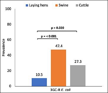
Figure 1. Prevalence of third-generation cephalosporin-resistant Escherichia coli (3GC-R E. coli) detected in fecal samples collected from laying hens, swine, cattle, and farm workers on a small-scale farm in northern Thailand. Bars represent the proportion of samples positive for 3GC-R E. coli within each host group. Statistically significant differences in prevalence among animal species were determined using Fisher’s exact test (p < 0.05).
Antibiotic susceptibility profiles
Assessment of the antibiotic susceptibility of 3GC-R E. coli against 12 antibiotics showed high resistance rates to β-lactam agents. Resistance to cefotaxime and cefpodoxime was detected in 100% of isolates, whereas resistance to ceftazidime and aztreonam was observed in 73.8% and 69.0% of isolates, respectively. Resistance to amoxicillin–clavulanate and cefepime ranged from 35.7% to 38.1% (Table 2).
| Antibiotics | Number of isolates (%) | ||||
|---|---|---|---|---|---|
|
| |||||
| Laying hens (n = 22) | Swine (n = 9) | Cattle (n = 9) | Farm workers (n = 2) | Total (95% CI) (n = 42) | |
| β-lactam | |||||
| Amoxicillin–Clavulanate | 12 (54.5) | 2 (22.2) | 1 (11.1) | 0 | 15 (35.7) (21.6–52.0) |
| Aztreonam | 13 (59.0) | 6 (66.7) | 8 (88.9) | 2 (100) | 29 (69.0) (52.9–82.4) |
| Cefepime | 7 (31.8) | 4 (44.4) | 3 (33.3) | 2 (100) | 16 (38.1) (23.6–54.4) |
| Cefotaxime | 22 (100) | 9 (100) | 9 (100) | 2 (100) | 42 (100) (93.1; 100) |
| Cefpodoxime | 22 (100) | 9 (100) | 9 (100) | 2 (100) | 42 (100) (93.1; 100) |
| Ceftazidime | 19 (86.4) | 5 (55.6) | 5 (55.6) | 2 (100) | 31 (73.8) (58.0–86.1) |
| Imipenem | 0 | 0 | 0 | 0 | 0 |
| Non β-lactam | |||||
| Chloramphenicol | 3 (13.6) | 6 (66.7) | 7 (77.8) | 1 (50.0) | 17 (40.5) (25.6–56.7) |
| Ciprofloxacin | 2 (9.1) | 3 (33.3) | 0 | 0 | 5 (11.9; 4.0–25.6) |
| Doxycycline | 6 (27.3) | 7 (77.8) | 7 (77.8) | 1 (50.0) | 21 (50.0) (34.2–65.8) |
| Gentamicin | 7 (31.8) | 6 (66.7) | 7 (77.8) | 1 (50.0) | 21 (50.0) (34.2–65.8) |
| Trimethoprim + Sulfamethoxazole | 9 (40.9) | 6 (66.7) | 5 (55.6) | 2 (100) | 22 (52.4) (36.4–68.0) |
| No. of antibiotics to which isolates are resistant | |||||
| 1–3 | 2 (9.1) | 0 | 0 | 0 | 2 (4.8) (0.6–16.2) |
| 4–6 | 12 (54.5) | 5 (55.6) | 4 (44.4) | 1 (50.0) | 22 (52.4) (36.4–68.0) |
| 7–9 | 8 (36.4) | 3 (33.3) | 5 (55.6) | 1 (50.0) | 17 (40.5) (25.6–56.7) |
| 10 | 0 | 1 (11.1) | 0 | 0 | 1 (2.4) (0.1–12.6) |
| Non-MDR | 13 (59.1) | 2 (22.2) | 2 (22.2) | 1 (50.0) | 18 (42.9) (27.8–59.0) |
| MDR | |||||
| 3 | 3 (13.6) | 1 (11.1) | 0 | 0 | 4 (9.5) (2.7–22.6) |
| 4 | 4 (18.2) | 1 (11.1) | 4 (44.4) | 0 | 9 (21.4) (10.3–36.8) |
| 5 | 1 (4.5) | 3 (33.3) | 3 (33.3) | 1 (50.0) | 8 (19.0) (8.6–34.1) |
| 6 | 1 (4.5) | 2 (22.2) | 0 | 0 | 3 (7.1) (1.5–19.5) |
| Total MDR | 9 (40.9) | 7 (77.8) | 7 (77.8) | 1 (50.0) | 24 (57.1) (41.0–72.3) |
Table 2. Antibiotic resistance profiles and MDR patterns of 3GC-R Escherichia coli isolated from farm animals and farm workers.
| Antibiotics | Number of isolates (%) | ||||
|---|---|---|---|---|---|
|
| |||||
| Laying hens (n = 22) | Swine (n = 9) | Cattle (n = 9) | Farm workers (n = 2) | Total (95% CI) (n = 42) | |
| β-lactam | |||||
| Amoxicillin–Clavulanate | 12 (54.5) | 2 (22.2) | 1 (11.1) | 0 | 15 (35.7) (21.6–52.0) |
| Aztreonam | 13 (59.0) | 6 (66.7) | 8 (88.9) | 2 (100) | 29 (69.0) (52.9–82.4) |
| Cefepime | 7 (31.8) | 4 (44.4) | 3 (33.3) | 2 (100) | 16 (38.1) (23.6–54.4) |
| Cefotaxime | 22 (100) | 9 (100) | 9 (100) | 2 (100) | 42 (100) (93.1; 100) |
| Cefpodoxime | 22 (100) | 9 (100) | 9 (100) | 2 (100) | 42 (100) (93.1; 100) |
| Ceftazidime | 19 (86.4) | 5 (55.6) | 5 (55.6) | 2 (100) | 31 (73.8) (58.0–86.1) |
| Imipenem | 0 | 0 | 0 | 0 | 0 |
| Non β-lactam | |||||
| Chloramphenicol | 3 (13.6) | 6 (66.7) | 7 (77.8) | 1 (50.0) | 17 (40.5) (25.6–56.7) |
| Ciprofloxacin | 2 (9.1) | 3 (33.3) | 0 | 0 | 5 (11.9; 4.0–25.6) |
| Doxycycline | 6 (27.3) | 7 (77.8) | 7 (77.8) | 1 (50.0) | 21 (50.0) (34.2–65.8) |
| Gentamicin | 7 (31.8) | 6 (66.7) | 7 (77.8) | 1 (50.0) | 21 (50.0) (34.2–65.8) |
| Trimethoprim + Sulfamethoxazole | 9 (40.9) | 6 (66.7) | 5 (55.6) | 2 (100) | 22 (52.4) (36.4–68.0) |
| No. of antibiotics to which isolates are resistant | |||||
| 1–3 | 2 (9.1) | 0 | 0 | 0 | 2 (4.8) (0.6–16.2) |
| 4–6 | 12 (54.5) | 5 (55.6) | 4 (44.4) | 1 (50.0) | 22 (52.4) (36.4–68.0) |
| 7–9 | 8 (36.4) | 3 (33.3) | 5 (55.6) | 1 (50.0) | 17 (40.5) (25.6–56.7) |
| 10 | 0 | 1 (11.1) | 0 | 0 | 1 (2.4) (0.1–12.6) |
| Non-MDR | 13 (59.1) | 2 (22.2) | 2 (22.2) | 1 (50.0) | 18 (42.9) (27.8–59.0) |
| MDR | |||||
| 3 | 3 (13.6) | 1 (11.1) | 0 | 0 | 4 (9.5) (2.7–22.6) |
| 4 | 4 (18.2) | 1 (11.1) | 4 (44.4) | 0 | 9 (21.4) (10.3–36.8) |
| 5 | 1 (4.5) | 3 (33.3) | 3 (33.3) | 1 (50.0) | 8 (19.0) (8.6–34.1) |
| 6 | 1 (4.5) | 2 (22.2) | 0 | 0 | 3 (7.1) (1.5–19.5) |
| Total MDR | 9 (40.9) | 7 (77.8) | 7 (77.8) | 1 (50.0) | 24 (57.1) (41.0–72.3) |
3GC-R = Third-generation cephalosporin-resistant, MDR = Multidrug resistance (resistance to ≥3 antimicrobial classes), CI = Confidence interval. Intermediate results were interpreted as susceptible according to Clinical and Laboratory Standards Institute guidelines.
Among non-β-lactam antibiotics, resistance to chloramphenicol, gentamicin, doxycycline, and trimethoprim–sulfamethoxazole ranged from 40.5% to 52.4%. Although resistance to ciprofloxacin was detected in 11.9% of isolates, all isolates remained susceptible to imipenem. Notably, isolates recovered from swine and cattle exhibited significantly higher resistance rates to chloramphenicol, gentamicin, and doxycycline compared with those from laying hens (Figure 2).
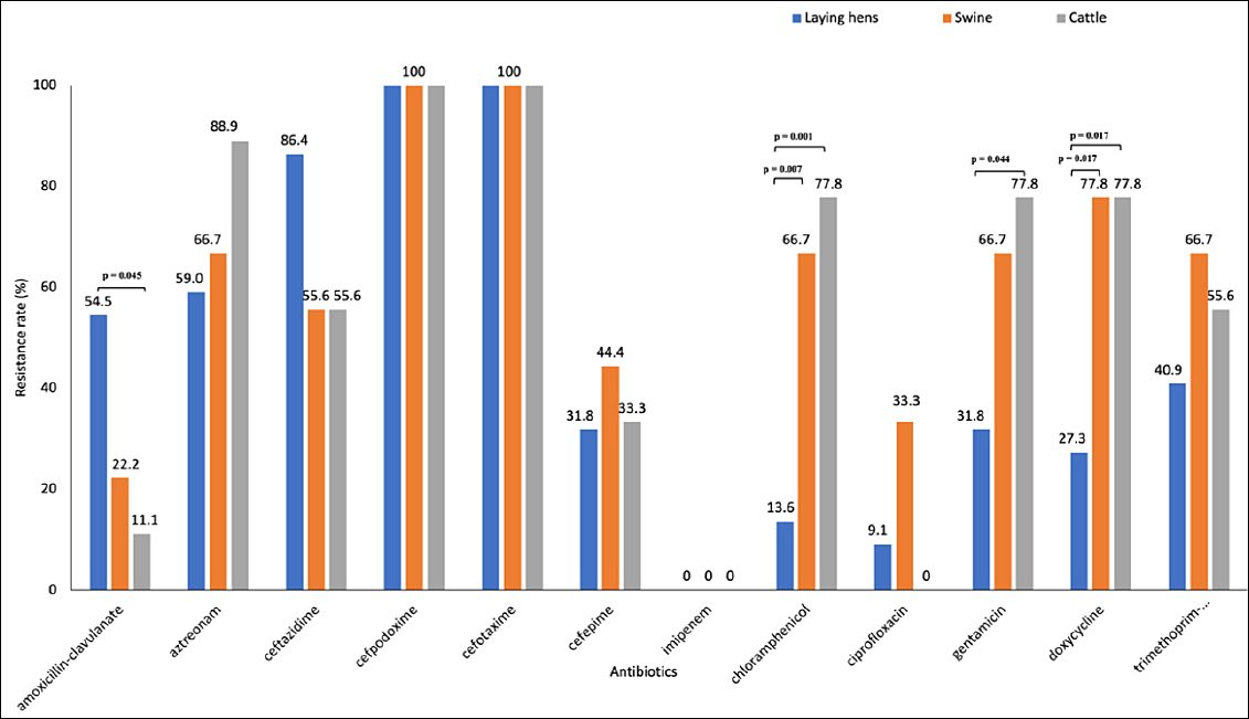
Figure 2. Antibiotic resistance rates of third-generation cephalosporin-resistant Escherichia coli isolates recovered from laying hens, swine, cattle, and farm workers. Resistance rates are expressed as percentages of isolates resistant to each tested antibiotic according to Clinical and Laboratory Standards Institute interpretive criteria. Differences in resistance profiles among host species highlight host-associated variation in antimicrobial resistance patterns.
MDR patterns
Most isolates (52.4%) exhibited resistance to 4–6 antibiotics, while 40.5% of isolates were resistant to 7–9 antibiotics (Table 2). A total of 24 distinct resistance profiles were identified. The most frequent resistance pattern was amoxicillin/clavulanate–ceftazidime–cefpodoxime–cefotaxime, which was detected in 14.3% of isolates (Table S3). Overall, 24 isolates (57.1%) were resistant to at least three antibiotic classes and were therefore classified as MDR. The largest proportion of isolates (21.4%) showed resistance to antibiotics belonging to four antimicrobial classes (Table 2).
Distribution of β-lactamase genes and ESBL production
Screening for β-lactamase genes demonstrated that all 3GC-R E. coli isolates harbored blaCTX-M, blaCMY-2, or both (Tables 1 and S2). Twenty-nine isolates (69.0%) carried blaCTX-M alone, including blaCTX-M-55 (n = 21), blaCTX-M-14 (n = 5), and blaCTX-M-15 (n = 3). Nine isolates (21.4%) simultaneously carried blaCTX-M and blaCMY-2. Among the 38 blaCTX-M-positive isolates, 28 (66.7%) were confirmed as ESBL producers (Tables 1 and S2). In addition, four isolates (9.5%) carried blaCMY-2 alone.
Transferability of resistance genes and plasmid replicon types
Conjugation experiments were performed using 23 3GC-R E. coli isolates as donors, including 19 blaCTX-M positive and four blaCMY-2 positive isolates. Of these, 22 isolates—comprising 11 blaCTX-M-55, five blaCTX-M-14, two blaCTX-M-15, and four blaCMY-2, successfully transferred their resistance genes to the recipient E. coli J53. Conjugation frequencies ranged from 4.1 × 10⁻⁸ to 7.2 × 10⁻² (Tables 3 and S4).
For transconjugants carrying either blaCTX-M or blaCMY-2, cefotaxime MIC values were increased by up to >32-fold compared with the recipient strain (E. coli J53; cefotaxime MIC = 0.25 µg/mL). Among these transconjugants, 13 were successfully characterized by PBRT. Detected plasmid replicon types included IncF (n = 5), IncFIA (n = 1), IncFIB (n = 3), and IncI1-Iγ (n = 4) in transconjugants carrying blaCTX-M and blaCMY-2 (Table 3).
| E. coli isolates carrying blaCTX-M or blaCMY-2 | Conjugation | ||
|---|---|---|---|
|
| |||
| Conjugation frequency a | Cefotaxime MIC (μg/mL) | Types of plasmid replicons (n) | |
| blaCTX-M-55 (n = 11) | 6.3 × 10⁻⁸– 7.2 × 10⁻² | 0.25 >8 b | IncF (n = 4) IncFIA (n = 1) Untypeable (n = 6) |
| blaCTX-M-14 (n = 5) | 4.1 × 10⁻⁸– 2.6 × 10⁻⁴ | 2– >8c | IncI1-Iγ (n = 2) IncFIB (n = 2) IncF (n = 1) |
| blaCTX-M-15 (n = 2) | 2.3 × 10⁻⁷– 5.8 × 10⁻⁷ | >8 | Untypeable (n = 2) |
| blaCMY-2 (n = 4) | 4.2 × 10⁻⁷– 1.3 × 10⁻³ | 0.25 >8 b | IncI1-Iγ (n = 2) IncFIB (n = 1) Untypeable (n = 1) |
Table 3. Transferability of blaCTX-M and blaCMY-2 genes in 3GC-R Escherichia coli and associated plasmid replicon types.
| E. coli isolates carrying blaCTX-M or blaCMY-2 | Conjugation | ||
|---|---|---|---|
|
| |||
| Conjugation frequency a | Cefotaxime MIC (μg/mL) | Types of plasmid replicons (n) | |
| blaCTX-M-55 (n = 11) | 6.3 × 10⁻⁸– 7.2 × 10⁻² | 0.25 >8 b | IncF (n = 4) IncFIA (n = 1) Untypeable (n = 6) |
| blaCTX-M-14 (n = 5) | 4.1 × 10⁻⁸– 2.6 × 10⁻⁴ | 2– >8c | IncI1-Iγ (n = 2) IncFIB (n = 2) IncF (n = 1) |
| blaCTX-M-15 (n = 2) | 2.3 × 10⁻⁷– 5.8 × 10⁻⁷ | >8 | Untypeable (n = 2) |
| blaCMY-2 (n = 4) | 4.2 × 10⁻⁷– 1.3 × 10⁻³ | 0.25 >8 b | IncI1-Iγ (n = 2) IncFIB (n = 1) Untypeable (n = 1) |
MIC = Minimum inhibitory concentration. a Conjugation frequency was calculated as the number of transconjugants divided by the number of donor cells. b One transconjugant showed a cefotaxime MIC of 0.25 µg/mL. c One transconjugant showed a cefotaxime MIC of 2 µg/mL. The MIC of cefotaxime for the recipient strain Escherichia coli J53 was 0.25 µg/mL.
Contribution of efflux pumps to ceftazidime resistance
To assess the role of efflux pumps in ceftazidime resistance, 31 3GC-R E. coli isolates resistant to ceftazidime by disk diffusion were further examined. According to CLSI guidelines [17], E. coli isolates with ceftazidime MICs of 8 µg/mL and ≥16 µg/mL are categorized as intermediate and fully resistant, respectively. Accordingly, ceftazidime MIC values ranged from 16 to 128 µg/mL in 29 isolates, while two isolates exhibited MICs of 8 µg/mL (Table S2).
In the presence of PAβN, four-fold and eight-fold reductions in ceftazidime MICs were observed in 11 and one isolates, respectively (Table 4). Inhibition of efflux pump activity by PAβN in a single isolate from cattle reduced the MIC from 8 µg/mL to 2 µg/mL (Table S2), restoring susceptibility to ceftazidime.
| Antibiotic (No. of tested isolates) | No. (%) of isolates with indicated fold reduction in MICs after addition of PAβN | ||||
|---|---|---|---|---|---|
|
| |||||
| No change | 2 | 4 | 8 | 16 | |
| Ceftazidime (n = 31) | 6 (19.4) | 13 (41.9) | 11 (35.5) | 1 (3.2) | 0 |
Table 4. Fold reduction in ceftazidime MICs of 3GC-R Escherichia coli in the presence of an efflux pump inhibitor.
| Antibiotic (No. of tested isolates) | No. (%) of isolates with indicated fold reduction in MICs after addition of PAβN | ||||
|---|---|---|---|---|---|
|
| |||||
| No change | 2 | 4 | 8 | 16 | |
| Ceftazidime (n = 31) | 6 (19.4) | 13 (41.9) | 11 (35.5) | 1 (3.2) | 0 |
MIC = Minimum inhibitory concentration, PAβN = Phenylalanine-arginine-β-naphthylamide. A reduction of ≥4-fold in MIC was considered indicative of efflux pump involvement.
Genetic relatedness of 3GC-R E. coli isolates
Genotypic analysis of 3GC-R E. coli isolates identified 23 distinct PFGE profiles (I–XXIII) among 41 isolates (Figure 3 and Table S2). One isolate from a farm worker could not be typed. Identical PFGE profiles were detected among isolates from laying hens (profiles I and II), swine (profile V), and cattle (profile VI). In addition, similarities were observed among isolates recovered from different animal species, with E. coli isolates from laying hens and swine sharing profiles III and IV, and isolates from swine and cattle sharing profile VI.
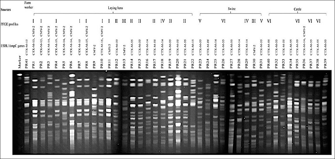
Figure 3. Pulsed-field gel electrophoresis (PFGE) patterns of cefotaxime-resistant Escherichia coli isolates obtained from laying hens, swine, cattle, and farm workers following XbaI digestion. PFGE profiles were analyzed to assess genetic relatedness among isolates from different host species. Interpretation of banding patterns was performed according to the criteria described by Tenover et al. [22]. Identical profiles indicate clonal relatedness, whereas distinct patterns reflect genetic diversity among isolates.
DISCUSSION
Prevalence of 3GC-R E. coli in small-scale livestock systems
The inappropriate use of antibiotics is a major driver of the emergence of ARB in livestock production systems. Previous studies conducted in low- and middle-income countries, particularly those focusing on small-scale farms, have reported a high prevalence of 3GC-R E. coli in farm animals, including chickens in Indonesia, Ecuador, and Zambia (66.1%–77.8%) [23–25], swine in Vietnam and Argentina (47.8%–89.0%) [26, 27], and cattle in Tanzania and Argentina (34.8%–95.0%) [26, 28]. Similarly, a recent study from small- and middle-scale farms in Peru reported ESBL-producing E. coli prevalences of 75.4% in dairy cows, 61.3% in pigs, and 34.1% in laying hens [29].
Overall, 3GC-R E. coli was detected in 15.8% of fecal samples collected from farm animals and workers (Table 1). This prevalence is lower than that reported in previous studies of food-producing animals raised on small-scale farms in Thailand (27.1%–55.6%) [15,16]. Differences in detection rates may be attributed to variations in study location, sample collection strategies, and isolation methodologies, all of which can influence prevalence estimates.
Species-specific prevalence and human carriage
The highest prevalence of 3GC-R E. coli was observed in swine (47.4%), followed by cattle (27.3%) and laying hens (10.5%) (Table 1). This distribution is consistent with reports from other countries, including Laos, Peru, and Argentina [26, 29, 30]. The higher prevalence observed in swine may be explained by the fact that pig production systems worldwide are associated with greater antimicrobial use than other livestock sectors, leading to increased antibiotic exposure and selective pressure for resistant strains [31].
In addition to livestock, 3GC-R E. coli was detected in the intestinal tracts of two of the three farm workers. This finding is consistent with previous reports demonstrating a high rate of 3GC-R E. coli carriage among apparently healthy individuals in this region [15], underscoring the potential occupational exposure risk in small-scale farming environments.
AMR profiles and MDR
Antimicrobial susceptibility testing revealed concerning resistance patterns. At least half of the isolates were resistant to third-generation cephalosporins (cefpodoxime and ceftazidime), aztreonam, doxycycline, gentamicin, and trimethoprim–sulfamethoxazole (Table 2), all of which are considered critically important antibiotics for the treatment of bacterial infections. Moreover, 57.1% of isolates were classified as MDR, indicating a substantial risk of treatment failure should infections occur.
Distribution of ESBL and pAmpC genes
The presence of ESBL-producing E. coli in livestock represents a significant public health concern, particularly in Africa and Asia [32]. Among the 42 3GC-R E. coli isolates examined in this study, blaCTX-M was predominantly detected either alone (n = 29) or in combination with blaCMY-2 (n = 9), while ESBL production was phenotypically confirmed in 28 isolates (Table 1). The absence of ESBL activity in ten blaCTX-M positive isolates may be attributed to low-level gene expression [33]. In addition, four isolates carried blaCMY-2 alone.
The detection of blaCTX-M-14, blaCTX-M-15, blaCTX-M-55, and blaCMY-2 in farm animals aligns with findings from multiple regions worldwide [34–36], as these enzymes represent the most prevalent ESBL and plasmid-mediated AmpC β-lactamases globally [4, 37].
Public health relevance of pAmpC-positive E. coli in livestock
In Thailand, pAmpC-producing E. coli have been frequently reported in human clinical samples [37,38], whereas reports from farm animals remain limited. Only a small number of blaCMY-2 positive E. coli isolates have previously been identified in domestic and wild animals, including pigs [38]. The presence of blaCMY-2 alone or in combination with blaCTX-M in farm animals in the present study (Table 1) suggests that livestock may serve as an underrecognized reservoir for pAmpC-positive 3GC-R E. coli in this region.
Horizontal transfer of resistance genes and plasmid dissemination
Conjugation experiments demonstrated that blaCTX-M and blaCMY-2 genes carried by 3GC-R E. coli isolates were transferable, resulting in cefotaxime MIC increases of up to >32-fold in transconjugants. These genes were associated with IncF and IncI1-Iγ plasmids, respectively (Table 2), both of which are well known for their capacity to disseminate among Enterobacterales.
IncF plasmids are the most frequently reported plasmids in E. coli and commonly harbor multiple blaCTX-M variants. Previous studies have shown that conjugative transfer of blaCTX-M is predominantly mediated by IncF plasmids [39], consistent with findings from backyard poultry in this region [15]. Similarly, two blaCMY-2 genes in this study were located on IncI1-Iγ plasmids, which are narrow-host-range conjugative plasmids frequently detected in food animal–associated Enterobacterales [40]. On these plasmids, blaCMY-2 is often embedded within insertion sequences, particularly ISEcp1, facilitating gene mobilization.
The detection of transferable blaCMY-2 on IncI1-Iγ plasmids in farm animal isolates provides new evidence of regional plasmid dissemination potential. These plasmids often carry additional resistance determinants, including those conferring resistance to fluoroquinolones and aminoglycosides, contributing to the multidrug-resistant phenotype commonly observed in 3GC-R E. coli [41]. The presence of such transferable resistance genes in rural farming environments represents a significant public health concern due to frequent human–animal–environment interactions.
Role of efflux pumps in ceftazidime resistance
Efflux pump overexpression is an important contributor to resistance against multiple antibiotic classes. In this study, the contribution of efflux pumps to ceftazidime resistance was assessed using PAβN, a well-characterized inhibitor of RND efflux pumps. PAβN has been shown to reduce MICs of third-generation cephalosporins and carbapenems in several Gram-negative bacteria, including E. coli [42], Klebsiella pneumoniae [11], and P. aeruginosa [10].
A ≥4-fold reduction in ceftazidime MICs in the presence of PAβN was observed in 38.7% of the isolates (Table 4), indicating a contributory role of RND efflux pumps in ceftazidime resistance. Complete restoration of susceptibility in one isolate further supports efflux-mediated resistance. However, among the 12 isolates showing efflux pump overexpression, only one exhibited an MDR phenotype (Table S2), consistent with previous reports suggesting that efflux pump overexpression alone does not necessarily confer MDR. Instead, efflux-mediated resistance may progress to MDR following additional mutational events [43]. A minor (two-fold) reduction in MICs observed in 41.9% of isolates further suggests partial efflux involvement.
Genetic relatedness and transmission dynamics
Transmission of ARB between livestock and farm workers has been documented in Japan, Vietnam, and Thailand [15, 27, 36]. Although high genetic diversity was observed among 3GC-R E. coli isolates in the present study, identical PFGE profiles were detected within and between animal species (Figure 3, Table S1), indicating that clonal dissemination can occur among animals raised in close proximity. Cross-species sharing of identical PFGE patterns between laying hens and swine and between swine and cattle supports local micro-ecological transmission rather than purely vertical spread.
Despite frequent interactions between animals and workers, clonal transmission between livestock and farm workers was not observed. Given the lack of direct contact among animal species, environmental dissemination via drainage water or manure is a plausible transmission route [44]. The absence of direct animal-to-human clonal spread in this study contrasts with previous reports [15, 36] and suggests that horizontal gene transfer via highly transferable plasmids carrying blaCTX-M and blaCMY-2 (Table 3) may play a more dominant role in resistance dissemination.
Implications for One Health surveillance and policy
The WHO and other international agencies have developed global strategies to mitigate AMR. Thailand has implemented two National Action Plans on AMR (NAP-AMR 2017–2021 and 2023–2027) [45, 46], which emphasize reducing AMR-related morbidity and mortality across human, animal, and environmental sectors. Surveillance of AMR bacteria, including 3GC-R E. coli, within a One Health framework is a core strategy of both plans.
Small-scale family-owned farms dominate livestock production in Thailand and are essential to the national economy. However, close human–animal contact in these systems increases zoonotic risk. The findings of this study strongly support the NAP-AMR strategy advocating expanded One Health surveillance of 3GC-R E. coli across humans, animals, and the environment in smallholder farming systems.
Study limitations and future perspectives
This study has several limitations. Sampling was limited to a single farm with a relatively small sample size. Environmental samples, including feed, manure, water, and surrounding soil, were not collected, which may have provided further insight into transmission pathways. Longitudinal sampling was not performed, preventing assessment of temporal trends in 3GC-R E. coli prevalence. In addition, data on antibiotic usage within the farm were unavailable, limiting the ability to identify selective pressures driving resistance emergence.
Future studies should incorporate whole-genome sequencing to characterize mobile genetic elements and integrons associated with bla genes. Expanded genomic surveillance across humans, animals, and environmental sources on multiple smallholder farms is warranted. Strengthening biosecurity practices and maintaining antibiotic usage records at the farm-level are critical steps toward controlling the emergence and dissemination of ARB.
CONCLUSION
This study demonstrated that 3GC-R E. coli are present in a small-scale, multi-species farming system in lower northern Thailand. Overall, 3GC-R E. coli were detected in 15.8% of fecal samples collected from laying hens, swine, cattle, and farm workers, with the highest prevalence observed in swine, followed by cattle and laying hens. A substantial proportion of isolates exhibited MDR, with more than half classified as MDR. Molecular analyses revealed that resistance was predominantly mediated by blaCTX-M (notably blaCTX-M-55, blaCTX-M-14, and blaCTX-M-15) alone or in combination with blaCMY-2. Importantly, these resistance determinants were located on transferable IncF and IncI1-Iγ plasmids, resulting in markedly increased cefotaxime MICs in transconjugants. In addition to enzymatic mechanisms, efflux pump activity contributed to ceftazidime resistance in a subset of isolates, highlighting the multifactorial nature of resistance. Although high genetic diversity was observed, identical PFGE profiles among different animal species indicated local clonal dissemination within the farm environment.
The detection of transferable ESBL- and pAmpC-producing E. coli in small-scale livestock systems has important implications for animal health, food safety, and public health. The presence of mobile resistance genes on highly transmissible plasmids underscores the risk of rapid horizontal dissemination within farm ecosystems and potentially beyond the farm boundary. Smallholder farms, where animals and humans live in close proximity and biosecurity measures are often inconsistently applied, may represent critical yet under-surveilled reservoirs for AMR. These findings support the urgent need for strengthened antimicrobial stewardship, improved farm-level biosecurity, and integrated One Health surveillance that includes humans, animals, and the environment.
A major strength of this study lies in its integrated One Health approach, combining phenotypic resistance profiling, molecular characterization of β-lactamase genes, plasmid transferability assessment, efflux pump analysis, and genotypic relatedness within a single small-scale farm setting. The inclusion of multiple livestock species and farm workers allowed a comprehensive evaluation of resistance dynamics across host interfaces. Furthermore, the demonstration of both clonal spread and plasmid-mediated horizontal gene transfer provides valuable insight into the mechanisms driving the persistence and dissemination of 3GC-R E. coli in rural farming systems.
In conclusion, small-scale farms in Thailand can serve as reservoirs of multidrug-resistant, 3GC-R E. coli carrying highly transferable resistance determinants. The coexistence of enzymatic resistance, efflux pump activity, and mobile plasmids highlights the complexity of AMR ecology in livestock production systems. Expansion of One Health–based surveillance, strict implementation of biosecurity measures, and improved documentation and regulation of antibiotic use at the farm-level are essential to mitigate the further spread of 3GC-R E. coli within rural communities and beyond.
DATA AVAILABILITY
The supplementary and sequencing data can be made available from the corresponding author upon request.
AUTHORS’ CONTRIBUTIONS
UT: Methodology, fieldwork, experiment, data collection and curation, and statistical analysis and interpretation. PRN: Conceptualization and supervision, methodology, data analysis and interpretation, and original draft writing, reviewing, and editing. Both authors have read and approved the final manuscript.
COMPETING INTERESTS
The authors declare that they have no competing interests.
PUBLISHER’S NOTE
Veterinary World remains neutral with regard to jurisdictional claims in the published institutional affiliations.
ACKNOWLEDGMENTS
This work was supported by Naresuan University (NU) and the National Science Research and Innovation Fund (Grant No. R2566B042). We are grateful to the farm owner for participating in this study.
REFERENCES
- WHO Global Antimicrobial Resistance and Use Surveillance System (GLASS):global antibiotic resistance surveillance report 2025. 2025. Accessed2025 Nov 27. [Available from] | [Google Scholar]
- Global burden of bacterial antimicrobial resistance 1990–2021:a systematic analysis with forecasts to 2050.. Lancet 2024;404(10459):1199-1226. [Google Scholar] | [Crossref]
- Malik H, Singh R, Kaur S, Dhaka P, Bedi JS, Gill JPS, Gongal G. Review of antibiotic use and resistance in food animal production in WHO South-East Asia Region. J Infect Public Health 2023;16(Suppl 1):172-182. [Google Scholar] | [Crossref]
- Poirel L, Madec JY, Lupo A, Schink AK, Kieffer N, Nordmann P, Schwarz S. Antimicrobial resistance in Escherichia coli. Microbiol Spectr 2018;6(4). [Google Scholar] | [Crossref]
- WHO bacterial priority pathogens list,2024:bacterial pathogens of public health importance to guide research, development and strategies to prevent and control antimicrobial resistance.. 2024. Accessed 2025 Aug 2. [Available from] | [Google Scholar]
- MacKinnon MC, McEwen SA, Pearl DL, Lyytikäinen O, Jacobsson G, Collignon P, Gregson DB, Valiquette L, Laupland KB. Mortality in Escherichia coli bloodstream infections:a multinational population-based cohort study. BMC Infect Dis 2021;21(1):606. [Google Scholar] | [Crossref]
- Tuamsuwan K, Chamawan P, Boonyarit P, Srisuphan V, Klaytong P, Rangsiwutisak C, Wannapinij P, Fongthong T, Stelling J, Turner P, Limmathurotsakul D. Frequency of antimicrobial-resistant bloodstream infections in 111 hospitals in Thailand, 2022. J Infect 2024;89(4):106249. [Google Scholar] | [Crossref]
- Al-Mustapha AI, Muasa B, Adetunji V, Awoyale OD, Adesiyan IM, Adesiji YO, Elelu N, Odetokun IA, Ogundijo OA, Yahaya R, Jibril AH, Awosanya EJ, Abubakar AT, Alimi Y, Ghali-Mohammed I, Raufu IA, Jimoh SM, Ezenduka E, Adetunji VO, Heikinheimo A. Global distribution and incidence of multidrug-resistant and ESBL-producing Escherichia coli: an observational study of the ATLAS dataset. Clin Epidemiol Glob Health 2025;35:102150. [Google Scholar] | [Crossref]
- Dulanto Chiang A, Dekker JP. Efflux pump-mediated resistance to new β-lactam antibiotics in multidrug-resistant Gram-negative bacteria. Commun Med (Lond) 2024;4(1):170. [Google Scholar] | [Crossref]
- Laudy AE, Osińska P, Namysłowska A, Zając O, Tyski S. Modification of the susceptibility of Gram-negative rods producing ESβLs to β-lactams by the efflux phenomenon. PLoS One 2015;10(3):e0119997. [Google Scholar] | [Crossref]
- Maurya N, Jangra M, Tambat R, Nandanwar H. Alliance of efflux pumps with β-lactamases in multidrug-resistant Klebsiella pneumoniae isolates. Microb Drug Resist 2019;25(8):1155-1163. [Google Scholar] | [Crossref]
- MacGowan AP, Attwood MLG, Noel AR, Barber R, Aron Z, Opperman TJ, Grimsey E, Stone J, Ricci V, Piddock LJV. Exposure of Escherichia coli to antibiotic-efflux pump inhibitor combinations in a pharmacokinetic model:impact on bacterial clearance and drug resistance. J Antimicrob Chemother 2023;78(12):2869-2877. [Google Scholar] | [Crossref]
- Percentage of susceptible organisms isolated from all specimens, 94 hospitals, Jan–Dec 2024. Accessed 2025 Nov 28. [Available from] | [Google Scholar]
- Tansawai U, Sanguansermsri D, Na-udom A, Walsh TR, Niumsup PR. Occurrence of extended-spectrum β-lactamase and AmpC genes among multidrug-resistant Escherichia coli and emergence of ST131 from poultry meat in Thailand. Food Control 2018;84:159-164. [Google Scholar] | [Crossref]
- Tansawai U, Walsh TR, Niumsup PR. Extended-spectrum β-lactamase-producing Escherichia coli among backyard poultry farms, farmers, and environments in Thailand. Poult Sci 2019;98(6):2622-2631. [Google Scholar] | [Crossref]
- Vannakovida C, Lampang KN, Chuammitri P, Punyapornwithaya V, Kreausukon K, Mektrirat R. Comparative occurrence and antibiogram of extended-spectrum β-lactamase-producing Escherichia coli among post-weaned calves and lactating cows from smallholder dairy farms in a parallel animal husbandry area. Vet World 2021;14(5):1311-1318. [Google Scholar] | [Crossref]
- Performance standards for antimicrobial susceptibility testing. Wayne (PA): CLSI; 2020. [Google Scholar]
- Falagas ME, Karageorgopoulos DE. Pandrug resistance (PDR), extensive drug resistance (XDR), and multidrug resistance (MDR) among Gram-negative bacilli:need for international harmonization in terminology. Clin Infect Dis 2008;46(7):1121-1122. [Google Scholar] | [Crossref]
- Woodford N, Fagan EJ, Ellington MJ. Multiplex PCR for rapid detection of genes encoding CTX-M extended-spectrum β-lactamases. J Antimicrob Chemother 2006;57(1):154-155. [Google Scholar] | [Crossref]
- Pérez-Pérez FJ, Hanson ND. Detection of plasmid-mediated AmpC β-lactamase genes in clinical isolates by using multiplex PCR. J Clin Microbiol 2002;40(6):2153-2162. [Google Scholar] | [Crossref]
- Carattoli A, Bertini A, Villa L, Falbo V, Hopkins KL, Threlfall EJ. Identification of plasmids by PCR-based replicon typing. J Microbiol Methods 2005;63(3):219-228. [Google Scholar] | [Crossref]
- Tenover FC, Arbeit RD, Goering RV, Mickelsen PA, Murray BE, Persing DH, Swaminathan B. Interpreting chromosomal DNA restriction patterns produced by pulsed-field gel electrophoresis:criteria for bacterial strain typing. J Clin Microbiol 1995;33(9):2233-2239. [Google Scholar] | [Crossref]
- Handayani KS, Setiyono A, Lukman DW, Pisestyani H, Rahayu P. Distribution of extended-spectrum β-lactamase-producing Escherichia coli genes in an integrated poultry-fish farming system in Bogor, Indonesia. Vet World 2024;17(7):1596-1602. [Google Scholar] | [Crossref]
- Hedman HD, Eisenberg JNS, Vasco KA, Blair CN, Trueba G, Berrocal VJ, Zhang L. High prevalence of extended-spectrum β-lactamase CTX-M-producing Escherichia coli in small-scale poultry farming in rural Ecuador. Am J Trop Med Hyg 2019;100(2):374-376. [Google Scholar] | [Crossref]
- Nasilele SJ, Shawa M, Kamboyi HK, Phiri BSJ, Chambaro H, Lundu T, Nundwe M, Lungu A, Moonga L, Kajihara M, Sawa H, Suzuki Y, Higashi H, Siame A, Mudenda NB, Hang'ombe MB, Muzandu K. Occurrence of cefotaxime-resistant and β-lactamase-producing Enterobacterales in poultry from small-scale farms in the Copperbelt province of Zambia. JAC Antimicrob Resist 2025;7(4):dlaf125. [Google Scholar] | [Crossref]
- Mounsey O, Marchetti L, Parada J, Alarcón LV, Aliverti F, Avison MB, Ayala CS, Ballesteros C, Best CM, Bettridge J, Buchamer A, Buldain D, Carranza A, Corti Isgro M, Demeritt D, Escobar MP, Gortari Castillo L, Jaureguiberry M, Lucas MF, Madoz LV, Marconi MJ, Moiso N, Nievas HD, Ramirez Montes De Oca MA, Reding C, Reyher KK, Vass L, Williams S, Giraudo J, De La Sota RL, Mestorino N, Moredo FA, Pellegrino M. Genomic epidemiology of third-generation cephalosporin-resistant Escherichia coli from Argentinian pig and dairy farms reveals animal-specific patterns of co-resistance and resistance mechanisms. Appl Environ Microbiol 2024;90(3):e01791-23. [Google Scholar] | [Crossref]
- Dang STT, Bortolaia V, Tran NT, Le HQ, Dalsgaard A. Cephalosporin-resistant Escherichia coliisolated from farm workers and pigs in northern Vietnam. Trop Med Int Health 2018;23(4):415-424. [Google Scholar] | [Crossref]
- Azabo RR, Mshana SE, Matee MI, Kimera SI. Antimicrobial resistance pattern of Escherichia coliisolates from small-scale dairy cattle in Dar es Salaam, Tanzania. Animals (Basel) 2022;12(14):1853. [Google Scholar] | [Crossref]
- Salgado-Caxito M, Léon D, Bardales O, Jara LM, Medrano P, Murga C, Pérez V, Aylas-Jurado B, Su-Tello R, Najarro J, Salvador-Tasayco E, Farrugia-Audri J, Shiva C, Benavides JA. Unexplained high prevalence of ESBL-Escherichia coli among cattle and pigs in Peru. Antibiotics (Basel) 2025;14(9):867. [Google Scholar] | [Crossref]
- Inthavong P, Chanthavong S, Nammanininh P, Phommachanh P, Theppangna W, Agunos A, Wagenaar JA, Douangngeun B, Loth L. Antimicrobial resistance surveillance of pigs and chickens in the Lao People's Democratic Republic, 2018–2021.. Antibiotics (Basel) 2022;11(2):177. [Google Scholar] | [Crossref]
- Thongyuan S, Tansakul N. Antimicrobial use on pig farms in Thailand:farmer perceptions of use and resistance. Prev Vet Med 2024;230:106287. [Google Scholar] | [Crossref]
- Mandujano-Hernández A, Martínez-Vázquez AV, Paz-González AD, Herrera-Mayorga V, Sánchez-Sánchez M, Lara-Ramírez EE, Vázquez K, de Jesús de Luna-Santillana E, Bocanegra-García V, Rivera G. The global rise of ESBL-producing Escherichia coli in the livestock sector:a five-year overview. Animals (Basel) 2024;14(17):2490. [Google Scholar] | [Crossref]
- Kjeldsen TSB, Overgaard M, Nielsen SS, Bortolaia V, Jelsbak L, Sommer M, Guardabassi L, Olsen JE. CTX-M-1 β-lactamase expression in Escherichia coli is dependent on cefotaxime concentration, growth phase and gene location. J Antimicrob Chemother 2015;70(1):62-70. [Google Scholar] | [Crossref]
- Chah KF, Ugwu IC, Okpala A, Adamu KY, Alonso CA, Ceballos S, Nwanta JN, Torres C. Detection and molecular characterization of extended-spectrum β-lactamase-producing enteric bacteria from pigs and chickens in Nsukka, Nigeria. J Glob Antimicrob Resist 2018;15:36-40. [Google Scholar] | [Crossref]
- Ewers C, de Jong A, Prenger-Berninghoff E, El Garch F, Leidner U, Tiwari SK, Semmler T. Genomic diversity and virulence potential of ESBL- and AmpC-β-lactamase-producing Escherichia coli strains from healthy food animals across Europe. Front Microbiol 2021;12:626774. [Google Scholar] | [Crossref]
- Nakano R, Nakano A, Nishisouzu R, Hikosaka K, Suzuki Y, Kamoshida G, Tansho-Nagakawa S, Endo S, Kasahara K, Ono Y, Yano H. Genetic relatedness of third-generation cephalosporin-resistant Escherichia coli among livestock, farmers, and patients in Japan. One Health 2023;16:100524. [Google Scholar] | [Crossref]
- Roberts T, Ling CL, Watthanaworawit W, Cheav C, Sengduangphachanh A, Silisouk J, Hopkins J, Phommasone K, Batty EM, Turner P, Ashley EA. AmpC β-lactamases detected in Southeast Asian Escherichia coli and Klebsiella pneumoniae. JAC Antimicrob Resist 2024;6(6):dla.e195. [Google Scholar] | [Crossref]
- Trongjit S, Assavacheep P, Samngamnim S, My TH, An VTT, Simjee S, Chuanchuen R. Plasmid-mediated colistin resistance and ESBL production in Escherichia coli from clinically healthy and sick pigs. Sci Rep 2022;12(1):2466. [Google Scholar] | [Crossref]
- Yu K, Huang Z, Xiao Y, Gao H, Bai X, Wang D. Global spread characteristics of CTX-M-type extended-spectrum β-lactamases:a genomic epidemiology analysis. Drug Resist Updat 2024;73:101036. [Google Scholar] | [Crossref]
- Foley SL, Kaldhone PR, Ricke SC, Han J. Incompatibility group I1 (IncI1) plasmids:their genetics, biology, and public health relevance. Microbiol Mol Biol Rev 2021;85(2):e00031-20. [Google Scholar] | [Crossref]
- Rozwandowicz M, Brouwer MSM, Fischer J, Wagenaar JA, Gonzalez-Zorn B, Guerra B, Mevius DJ, Hordijk J. Plasmids carrying antimicrobial resistance genes in Enterobacteriaceae. J Antimicrob Chemother 2018;73(5):1121-1137. [Google Scholar] | [Crossref]
- Rodríguez-Villodres Á, Ortiz de la Rosa JM, Gálvez-Benítez L, Gascón ML, Peñalva G, Dorado Pardo FJ, Casimiro-Soriguer CS, Rodríguez Rodríguez M, Amaya Villar R, Cisneros JM, Lepe JA. Survival of infection with TEM β-lactamase-producing Escherichia coli with pan-β-lactam resistance. J Infect 2024;89(5):106268. [Google Scholar] | [Crossref]
- Ebbensgaard AE, Løbner-Olesen A, Frimodt-Møller J. The role of efflux pumps in the transition from low-level to clinical antibiotic resistance. Antibiotics (Basel) 2020;9(12):855. [Google Scholar] | [Crossref]
- Graham DW, Bergeron G, Bourassa MW, Dickson J, Gomes F, Howe A, Kahn LH, Morley PS, Scott HM, Simjee S, Singer RS, Smith TC, Storrs C, Wittum TE. Complexities in understanding antimicrobial resistance across domesticated animal, human, and environmental systems. Ann N Y Acad Sci 2019;1441(1):17-30. [Google Scholar] | [Crossref]
- . Accessed 2025 Nov 27. [Available from] | [Google Scholar]
- . Accessed 2025 Nov 27. [Available from] | [Google Scholar]