

ABSTRACT
Background and Aim: Antimicrobial resistance (AMR) in extended-spectrum β-lactamase (ESBL)–producing
Materials and Methods: A cross-sectional surveillance study was conducted from May to August 2025 on 59 dairy farms. One sample per matrix per farm was collected, including milk, forage, soil, animal drinking water, hand-wash water, and feces (total n = 354). Isolation and phenotypic identification of
Results:
Conclusion: The widespread detection of MDR and ESBL-producing
Keywords: antimicrobial resistance, dairy farm, extended-spectrum beta-lactamase, Indonesia,
INTRODUCTION
Dairy cattle farming is vital to Indonesia’s food system, making the health of animals and milk quality crucial for producing safe, high-quality food for people [1, 2]. East Java is among the top provinces in the country for dairy cattle numbers and milk output, with Batu City contributing significantly by producing about 25,258 liters of milk daily [2, 3]. Many dairy farms in Batu City also serve as educational agrotourism spots, fostering frequent interactions among livestock, farm settings, and the public. These public-facing farms may increase exposure to antimicrobial-resistant (AMR) bacteria, positioning dairy farms as key points in the spread of antimicrobial resistance within a One Health framework. The detection of multidrug-resistant (MDR) and extended-spectrum β-lactamase (ESBL)-producing
The quality of milk depends heavily on proper herd health management and disease control practices, which often involve the use of antibiotics for treatment and prevention [4, 5]. However, the widespread and uncontrolled use of antibiotics in dairy farming has led to the global rise of antimicrobial resistance, reducing treatment effectiveness, extending disease duration, and increasing the spread of antibiotic residues and resistance genes in the environment [6–8]. According to the Global Research on Antimicrobial Resistance report, infections caused by resistant bacteria resulted in about 4.95 million deaths worldwide in 2019, with forecasts pointing to significant impacts on global health and livestock production by 2050 [9, 10]. A key mechanism behind antimicrobial resistance is the production of enzymes that break down β-lactam antibiotics, making bacteria resistant to third-generation cephalosporins and similar drugs [11, 12]. ESBL production is often linked with MDR, as ESBL-producing bacteria frequently show resistance to other antibiotic classes, including aminoglycosides and chloramphenicol [13, 14]. Among Enterobacteriaceae,
Despite rising reports of antimicrobial resistance in livestock systems, most studies in Indonesia have concentrated on clinical isolates, milk samples, or wastewater surveillance, with limited focus on the broader farm ecosystem where resistant bacteria may persist and circulate. Dairy farms are complex environments where animals, feed, water, soil, equipment, and human-contact surfaces continually interact, creating multiple opportunities for resistant bacteria to be maintained and spread. However, data on the occurrence of multidrug-resistant and extended-spectrum β-lactamase-producing
Therefore, the present study was carried out to conduct a comprehensive multimatrix surveillance of antimicrobial resistance, multidrug resistance, and extended-spectrum β-lactamase-producing
MATERIALS AND METHODS
Ethical approval
Ethical approval for this study was obtained from the Health Research Ethics Committee (Komite Etik Penelitian Kesehatan, KEPK), Universitas Muhammadiyah Malang, Indonesia, under approval number E.5.a/065/KEPKUMM/IV/2025. Before sample collection, permission to conduct the study was obtained from the relevant local authority in Batu City and from the owners or managers of all participating dairy farms. Written or verbal informed consent was obtained from farm owners or responsible personnel before enrollment in the study.
All sampling procedures were performed using non-invasive methods and in accordance with accepted animal welfare principles. Milk samples were collected during routine milking procedures after standard udder hygiene practices, whereas fecal samples were collected as freshly voided material from the ground without restraining or disturbing the animals. Environmental samples, including forage feed, soil, animal drinking water, and hand-wash water, were collected without causing harm, stress, or disruption to the animals or farm activities. No experimental infection, invasive handling, or therapeutic intervention was performed on the animals as part of this study.
All field and laboratory procedures were carried out in accordance with biosafety and biosecurity measures to minimize contamination risks and protect animals, farm workers, and researchers. The confidentiality of farm identity and participant information was maintained throughout the study, and the collected data were used solely for research purposes.
Study design, period, and location
This study used a cross-sectional, surveillance-based approach to examine the presence of MDR and ESBL-producing
The study focused on farm-level environmental surveillance rather than investigating clinical cases, with sampling designed to capture multiple on-farm matrixes representing animal-derived products, environmental reservoirs, and human-associated interfaces. This approach was adopted to support a One Health surveillance framework by assessing potential AMR reservoirs and transmission pathways within dairy production systems before environmental dissemination occurs downstream.
Farm selection, eligibility criteria, and sample size
During the study period, dairy farms were chosen based on convenience and voluntary participation. Farms in Batu City were identified through local dairy farmer associations and cooperative networks and invited to join the study. A total of 59 dairy farms agreed to participate and were included.
Farms were eligible for inclusion if they: (i) were actively producing raw cow’s milk during the study period; (ii) operated as small- to medium-sized dairy farms; (iii) allowed access for milk and environmental sample collection; and (iv) obtained informed consent from farm owners or responsible workers. Farms were excluded if they were not in active production, declined participation, or did not permit comprehensive sampling across the targeted matrixes.
A single-proportion formula was used to estimate the sample size for cross-sectional prevalence studies. Assuming an expected ESBL prevalence of 11% [29], a 95% confidence level (Z = 1.96), and a precision of 10%, the minimum required sample size was calculated to be 38 farms. The inclusion of 59 dairy farms exceeded this minimum requirement and was considered sufficient to generate baseline surveillance data for
Sample collection, transport, and storage
To reduce contamination and ensure reproducibility across farms, sample collection was performed using standardized protocols. Samples were collected once per farm during the study period, with one sample taken for each predefined matrix per farm to broadly represent potential AMR within each dairy farm ecosystem. This method was chosen to support baseline surveillance at the farm-level rather than within-farm quantitative comparisons. To prevent cross-contamination, sterile tools were used for each sample, disposable gloves were changed between different sample types, and all samples were handled aseptically during collection and transport.
Milk samples (30 mL) were collected from individual lactating cows during routine milking after discarding the initial streams of milk and following standard udder hygiene procedures. The samples were stored in sterile containers. Animal drinking water samples (1,000 mL) were collected directly from water troughs used by the cattle, while hand-washing water samples (200 mL) were taken from designated hand-washing stations after milking to represent human-associated environmental exposure on the farm.
Forage feed samples (200 g) were aseptically collected from feed bunks or feed storage areas commonly accessed by cattle. Soil samples (25 g) were taken from areas around cattle housing at an approximate depth of 10 cm and within 1–2 m of the barn to represent the immediate farm environment. Freshly voided fecal samples (10 g) were gathered from the ground to minimize environmental contamination.
Six sample types were collected from each farm, resulting in 354 samples from 59 participating farms. All samples were immediately placed in sterile containers, stored in insulated cool boxes with ice packs, and transported under cold-chain conditions. The samples’ temperatures were monitored using calibrated digital thermometers inside the cool boxes, with readings recorded at the start of transport and upon arrival at the lab to ensure they stayed at approximately 4°C throughout transportation [30]. All samples were delivered to the Veterinary Microbiology and Immunology Laboratory, Faculty of Veterinary Medicine, Universitas Brawijaya, and processed within 6–12 h of collection. Sampling was conducted after obtaining permission from the relevant farm owners and local authorities.
Isolation and phenotypic identification of K. pneumoniae strains
All collected samples were inoculated into Buffered Peptone Water (BPW) (Oxoid, Basingstoke, UK) at a 1:9 ratio and then incubated at 37°C for 24 h. Samples were pre-enriched in BPW with a 1:9 sample-to-broth ratio to improve the recovery of stressed or low-abundance bacteria and to neutralize any inhibitory substances in environmental and milk matrixes. BPW was chosen as a non-selective pre-enrichment medium according to ISO (International Organization for Standardization) and FDA BAM (Food and Drug Administration Bacteriological Analytical Manual) protocols [31].
The BPW isolates were then cultured on MacConkey Agar (MCA) (Oxoid). Colonies displaying mucoid morphology, presumed to be

Figure 1. Isolation and identification of

Figure 2. Identification results of
Antimicrobial susceptibility test results
Pure
Antibiotic disks were placed on the agar surface with a center-to-center distance of approximately 25–30 mm, followed by incubation at 35°C ± 2°C for 16–18 h (Figure 3) [35]. Antimicrobial susceptibility testing was performed according to the 2020 Clinical and Laboratory Standards Institute (CLSI) guidelines, including standardized inoculum preparation adjusted to the 0.5 McFarland turbidity standard and controlled incubation conditions.

Figure 3. Confirmatory test of antibiotic sensitivity by disk diffusion with the Kirby–Bauer method on Mueller–Hinton agar media.
Although a reference quality-control strain (e.g.,
ESBL confirmation test
The double-disk synergy test (DDST) is a phenotypic assay used to identify
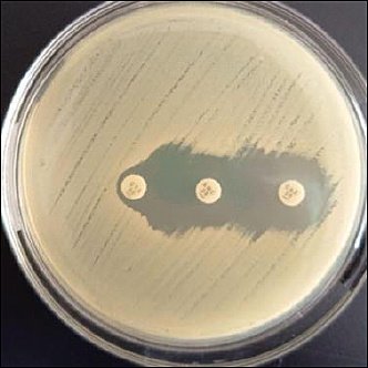
Figure 4. Positive results of extended-spectrum β-lactamase-producing bacteria on the double-disk synergy test.
Quality assurance
Quality assurance procedures were followed during sample collection, transportation, and laboratory analysis to ensure data accuracy and consistency in methods. All sampling equipment and laboratory consumables were sterilized and used only once per sample to prevent cross-contamination. Culture media and reagents were prepared according to the manufacturer’s instructions and ISO and FDA BAM guidelines. Media sterility was confirmed by incubating uninoculated control plates alongside test samples.
Laboratory instruments, including incubators and refrigerators, were routinely calibrated and monitored to ensure proper functioning. Bacterial suspensions used for antimicrobial susceptibility testing were standardized with a 0.5 McFarland turbidity standard. Quality control for antimicrobial susceptibility testing was conducted according to CLSI guidelines. A randomly selected subset of isolates was re-tested to confirm reproducibility. All laboratory procedures were performed by trained personnel following standardized operating procedures. Data entry and laboratory records were cross-checked to reduce transcription errors and ensure consistency between laboratory results and analytical datasets.
Statistical analysis
Data were analyzed descriptively. Prevalence estimates were calculated as proportions and presented with 95% confidence intervals. Binomial proportion confidence intervals were calculated to quantify the precision of the prevalence estimates. Statistical analyses were performed using Microsoft Excel version 21 (Microsoft Corp., Redmond, WA, USA).
RESULTS
Isolation and phenotypic identification of K. pneumoniae strains
Isolation and phenotypic identification of samples collected from 59 dairy farms in Batu City showed that
Table 1. Identification of
| Sample type | Total samples (n) | Positive n (%) | 95% Confidence interval |
|---|---|---|---|
| Hand-wash water | 59 | 42 (71.2) | 59.1–81.2 |
| Animal drinking water | 59 | 44 (74.6) | 62.9–83.8 |
| Milk | 59 | 30 (50.8) | 37.6–64.0 |
| Soil | 59 | 42 (71.2) | 59.1–81.2 |
| Forage feed | 59 | 50 (84.7) | 73.5–92.4 |
| Feces | 59 | 9 (15.3) | 7.3–27.0 |
| Total | 354 | 217 (61.3) | 56.1–66.3 |
Antibiotic susceptibility test results
Antimicrobial susceptibility testing of
Table 2. Antimicrobial resistance patterns of
| AMR data | Hand-wash water (n = 42) | Animal drinking water (n = 44) | Milk (n = 30) | Soil (n = 42) | Forage feed (n = 50) | Feces (n = 9) | Subtotal (n = 217) |
|---|---|---|---|---|---|---|---|
| CTX | |||||||
| R | 17 (40.5) | 15 (34.1) | 12 (40) | 14 (33.3) | 23 (46) | 2 (22.2) | 83 (38.3) |
| I | 7 (16.7) | 9 (20.4) | 4 (13.3) | 11 (26.2) | 11 (22) | 1 (11.1) | 43 (19.8) |
| S | 18 (42.8) | 20 (45.5) | 14 (46.67) | 17 (40.5) | 16 (32) | 6 (66.7) | 91 (41.9) |
| C | |||||||
| R | 3 (7.1) | 3 (6.8) | 7 (23.33) | 6 (14.3) | 8 (16) | 0 (0) | 27 (12.5) |
| I | 7 (16.7) | 5 (11.4) | 1 (3.33) | 3 (7.1) | 5 (10) | 2 (22.2) | 23 (10.6) |
| S | 32 (76.2) | 36 (81.8) | 22 (73.33) | 33 (78.6) | 37 (74) | 7 (77.8) | 167 (76.9) |
| AMP | |||||||
| R | 37 (88.1) | 42 (95.5) | 25 (83.33) | 37 (88.1) | 45 (90) | 8 (88.9) | 194 (89.4) |
| I | 5 (11.9) | 2 (4.5) | 3 (10) | 1 (2.4) | 3 (6) | 1 (11.1) | 15 (6.9) |
| S | 0 (0) | 0 (0) | 2 (6.66) | 4 (9.5) | 2 (4) | 0 (0) | 8 (3.7) |
| TE | |||||||
| R | 3 (7.1) | 5 (11.4) | 5 (16.7) | 3 (7.1) | 4 (8) | 0 (0) | 20 (9.2) |
| I | 1 (2.4) | 1 (2.2) | 0 (0) | 0 (0) | 1 (2) | 0 (0) | 3 (1.4) |
| S | 38 (90.5) | 38 (83.4) | 25 (83.33) | 39 (92.9) | 45 (90) | 9 (100) | 194 (84.4) |
| S | |||||||
| R | 25 (59.5) | 31 (70.5) | 22 (73.33) | 31 (73.8) | 38 (76) | 8 (88.9) | 155 (71.4) |
| I | 11 (26.2) | 11 (25) | 6 (20) | 7 (16.7) | 9 (18) | 1 (11.1) | 45 (20.8) |
| S | 6 (14.3) | 2 (4.5) | 2 (6.66) | 4 (9.5) | 3 (6) | 0 (0) | 17 (7.8) |
| SXT | |||||||
| R | 6 (14.3) | 5 (11.4) | 5 (16.7) | 6 (14.3) | 4 (8) | 1 (11.1) | 27 (12.5) |
| I | 2 (4.8) | 2 (4.5) | 1 (3.33) | 5 (11.9) | 0 (0) | 0 (0) | 10 (4.6) |
| S | 34 (80.9) | 37 (84.1) | 24 (80) | 31 (73.8) | 46 (92) | 8 (88.9) | 180 (82.9) |
| CIP | |||||||
| R | 1 (2.4) | 0 (0) | 0 (0) | 2 (4.8) | 3 (6) | 0 (0) | 6 (2.8) |
| I | 1 (2.4) | 2 (4.5) | 1 (3.33) | 0 (0) | 1 (2) | 0 (0) | 5 (2.3) |
| S | 40 (95.2) | 42 (95.5) | 29 (96.67) | 40 (95.2) | 46 (92) | 9 (100) | 206 (94.9) |
AMP = Ampicillin, AMR = Antimicrobial resistance, C = Chloramphenicol, CIP = Ciprofloxacin, CTX = Cefotaxime, I = Intermediate, R = Resistant, S = Streptomycin, S = Susceptible, SXT = Sulfamethoxazole–trimethoprim, TE = Tetracycline.
Isolates resistant to three or more classes of antimicrobial agents were classified as MDR [38]. Among the 217
Table 3. Distribution of MDR
| Antimicrobial classes | Hand-wash water | Animal drinking water | Milk | Soil | Forage feed | Feces | Subtotal n (%) |
|---|---|---|---|---|---|---|---|
| 3 | 13 (27.7) | 10 (21.3) | 5 (10.5) | 6 (12.8) | 11 (23.4) | 2 (4.3) | 47 (61.8) |
| 4 | 0 (0.0) | 4 (36.4) | 2 (18.2) | 0 (0.0) | 5 (45.4) | 0 (0.0) | 11 (14.5) |
| 5 | 2 (12.5) | 1 (6.3) | 2 (12.5) | 7 (43.7) | 4 (25.0) | 0 (0.0) | 16 (21.1) |
| 6 | 0 (0.0) | 0 (0.0) | 2 (100) | 0 (0.0) | 0 (0.0) | 0 (0.0) | 2 (2.6) |
| 7 | 0 (0.0) | 0 (0.0) | 0 (0.0) | 0 (0.0) | 0 (0.0) | 0 (0.0) | 0 (0.0) |
| Total | 76 (100) |
MDR = Multidrug-resistant.
MDR was defined as resistance to three or more classes of antimicrobial agents. Percentages within rows represent the distribution of MDR isolates across sample types for each resistance category. Subtotal percentages represent the overall distribution of MDR isolates. Percentages were calculated using the total number of MDR isolates (n = 76) as the denominator. Values 3–7 indicate the number of different antimicrobial classes to which isolates exhibit resistance.
ESBL confirmation test
Among the 76 MDR
Table 4. Prevalence of ESBL-producing
| Sample type | ESBL-positive (n) | Prevalence (%) | 95% CI |
|---|---|---|---|
| Hand-wash water | 1 | 1.3 | 0.2–7.1 |
| Animal drinking water | 5 | 6.6 | 2.9–14.5 |
| Milk | 1 | 1.3 | 0.2–7.1 |
| Soil | 3 | 3.9 | 1.3–10.8 |
| Forage feed | 6 | 7.9 | 3.7–15.9 |
| Feces | 0 | 0 | 0.0–4.8 |
| Total | 16 | 21.1 | 13.1–31.9 |
Prevalence was calculated based on the total number of MDR
DISCUSSION
Occurrence of K. pneumoniae in dairy farm matrixes
This study shows a high presence of
Comparison with previous studies and possible sources of contamination
Compared with previous studies conducted in the Republic of the Congo and China, which reported much lower
Antimicrobial resistance patterns in dairy farm isolates
Antimicrobial susceptibility testing showed high resistance levels to antibiotics commonly used in food-producing animals, especially ampicillin and streptomycin. These results agree with reports from Indonesia and other Southeast Asian nations, where penicillins, sulfonamides, tetracyclines, and quinolones are frequently used in livestock production [24]. Studies from Southeast Asia consistently indicate that extensive and sometimes inappropriate use of antimicrobials in dairy farming leads to higher resistance rates, particularly to β-lactam antibiotics. The similarity between the resistance patterns observed in this study and those reported in wastewater surveillance suggests that dairy farms serve as significant upstream sources of AMR, contributing to environmental contamination downstream [28]. However, unlike wastewater research, the current farm-level, multimatrix approach allows for earlier detection of resistance sources before they are diluted and dispersed.
Multidrug resistance and antimicrobial usage pressure
The proportion of MDR
Distribution of ESBL-producing isolates in environmental matrixes
The detection of ESBL-producing
Environmental persistence of ESBL-producing bacteria
The environmental persistence of ESBL-producing
Horizontal gene transfer and transmission risk within-farm ecosystems
Beyond persistence, the co-occurrence of MDR and ESBL-producing
One Health implications of MDR and ESBL dissemination
The concurrent detection of MDR- and ESBL-producing
Novel contributions and significance of the present study
Overall, this study offers several new contributions to research on AMR in dairy production systems. It provides a farm-level, multimatrix assessment of
Limitations and future research directions
This study has several limitations to consider when interpreting the results. First, the cross-sectional design offers a snapshot of AMR at a single point in time and cannot assess changes over time or seasonal variations in the occurrence and resistance patterns of
Third, the study was conducted in a single geographic area and involved one-time sampling per farm, which may limit the applicability of the findings to other dairy production systems with different management practices or environmental conditions. Additionally, although environmental and animal-associated matrixes were thoroughly assessed, human carriage was not evaluated, preventing direct conclusions about zoonotic transmission to farm workers or visitors. Furthermore, the study did not quantitatively analyze farm-level risk factors, such as hygiene practices, water source quality, or feed storage conditions, which may affect the distribution and persistence of AMR bacteria.
This study relied on phenotypic AMR testing following the CLSI guidelines; however, the lack of a reference quality-control strain in the AMR test (e.g.,
Despite these limitations, the current study provides valuable baseline data on the prevalence of MDR and ESBL-producing
CONCLUSION
This study showed a high presence of
From a practical perspective, the dominance of MDR and ESBL-producing
A key strength of this study is its multimatrix, farm-level surveillance method, which enabled the detection of upstream reservoirs of MDR and ESBL-producing bacteria that are often missed in clinical or wastewater studies. By analyzing milk, feces, feed, soil, drinking water, and hand-wash water simultaneously, this research offers a more comprehensive understanding of resistance spread within dairy farm environments and supports using a One Health approach for AMR surveillance.
In conclusion, dairy farm environments in Batu City may act as key reservoirs for MDR and ESBL-producing
DATA AVAILABILITY
The datasets generated and/or analyzed during the current study are available from the corresponding author upon reasonable request.
AUTHORS’ CONTRIBUTIONS
FNAEPD: Conceptualization, study design, supervision of laboratory work, critical review of the manuscript, and corresponding author responsibility. SMY: Study design, data interpretation, and manuscript revision. ENU: Antimicrobial resistance interpretation and critical revision of the manuscript. ARK: Antimicrobial resistance interpretation and manuscript revision. MHE: Study design, data interpretation, and critical revision of the manuscript. RS: Data analysis, data interpretation, drafting of the manuscript, and manuscript revision. MAAS: Sample collection and preliminary data processing. IAA, SK, and SLA: Supervision of laboratory work and critical review of the manuscript. TE, NYN, BAM, and NAPD: Field sampling, data entry, laboratory analyses, bacterial isolation, bacterial identification, and antimicrobial susceptibility testing. All authors have read and approved the final version of the manuscript.
COMPETING INTERESTS
The authors declare that they have no competing interests.
PUBLISHER’S NOTE
Veterinary World remains neutral with regard to jurisdictional claims in the published institutional affiliations.
ACKNOWLEDGMENTS
The authors would like to express their sincere gratitude to the Dairy Cooperative (KUD) of Batu City for granting permission and facilitating access to the dairy farms involved in this study. We also thank the participating dairy farmers for their cooperation and support during sample collection. Appreciation is extended to the staff of the Veterinary Microbiology and Immunology Laboratory, Faculty of Veterinary Medicine, Universitas Brawijaya, for their technical assistance and laboratory support throughout the study. This research was supported by the Penelitian Doktor Baru grant, Faculty of Veterinary Medicine, Universitas Brawijaya, under the DPP/SPP 2025 funding scheme (Dean’s Decree No. 30/2025).
REFERENCES
- Jahroh S, Atmakusuma J, Harmini, Fadillah A. Comparative Analysis of Dairy Farming Management and Business Model Between East Java and West Java, Indonesia. JMA [Internet] 2020;17((1)):96. [Google Scholar] | [Crossref]
- Kristanti ND, Fernandes LMY, Windari W, ShikhMaidin M. Milking Biosecurity in Dairy Cattle Farming East Java Indonesia. JAPSI [Internet] 2024. [cited 2026 Jan 12]. [Available from] | [Google Scholar]
- Yusuf H, Rukkwamsuk T, Idris S, Paul M. Antimicrobial usage surveillance of cattle in Indonesia to address antimicrobial resistance. ASSEHR 2018;98:355-359. [Google Scholar] | [Crossref]
- Harun MH, Shimelis S, Andargie B. Assessment of biosecurity status in dairy cow farms. Bull Natl Res Cent [Internet] 2022. [cited 2026 Jan 12]. [Available from] | [Google Scholar]
- Alabi MA, Chenia HY, Lin J. Antibiotic use in livestock: a driver of resistance in Africa and path to safer alternatives. MicrobiologyOpen 2025;14:e70122. [Google Scholar] | [Crossref]
- Mitima HI, Kawala LZ, Kajiramugabi JBI. Antimicrobial resistance in raw cow's milk in Bwegera, Democratic Republic of the Congo: implications for food safety. BMC Microbiol 2025;25:716. [Google Scholar] | [Crossref]
- Qiao M, Ying GG, Singer AC, Zhu YG. Review of antibiotic resistance in China and its environment. Environ Int 2018;110:160-172. [Google Scholar] | [Crossref]
- Ahmed SK, Hussein S, Qurbani K, Ibrahim RH, Fareeq A, Mahmood KA, Mohamed MG. Antimicrobial resistance: impacts, challenges, and future prospects. J Med Surg Public Health 2024;2:100081. [Google Scholar] | [Crossref]
- Pires AJ, Pereira G, Fangueiro D, Bexiga R, Oliveira M. When the solution becomes the problem: a review on antimicrobial resistance in dairy cattle. Future Microbiol 2024;19((10)):903-929. [Google Scholar] | [Crossref]
- Rahaman A, Mimi A, Antor MTH, Bakhtiyar Z, Hasan MAE, Fahim NAI, Jany DA, Rahman MT. Prevalence of extended-spectrum beta-lactamase-producing Enterobacteriaceae isolated from animals in Bangladesh: a systematic review and meta-analysis. One Health 2025;21:101237. [Google Scholar] | [Crossref]
- Rastuti MR, Budayanti NNS, Dwija IBNP. Various types of extended-spectrum β-lactamases: a literature review. J Clin Microbiol Infect Dis 2023;3((2)):29-34. [Google Scholar] | [Crossref]
- Zhang YL, Huang FY, Gan LL. High prevalence of
bla CTX-M andbla SHV among ESBL-producingEscherichia coli isolates from beef cattle in China's Sichuan–Chongqing Circle. Sci Rep 2021;11:13725. [Google Scholar] | [Crossref] - Prity SS, Akter K, Aktar MB, Mehzabin K, Jarin L, Shilpi RY. Multidrug-resistant ESBL-producing Enterobacteriaceae associated with clinical samples in a tertiary care hospital, Sirajganj. Eur J Med Health Sci 2024;6((1)):44-49. [Google Scholar] | [Crossref]
- Green LH, Goldman E. Practical handbook of microbiology. Abingdon: CRC Press; 2021. [Google Scholar]
- Parmar N, Singh R, Tyagi A. Antimicrobial resistance profiles and genomic insights of phenotypically extended-spectrum beta-lactamase-positive
K. pneumoniae from cattle farms. Curr Genet 2025;71:23. [Google Scholar] | [Crossref] - Kamaruzzaman EA, Aziz SA, Bitrus AA, Zakaria Z, Hassan L. Occurrence and characteristics of extended-spectrum beta-lactamase-producing
Escherichia coli from dairy cattle, milk, and farm environments in Peninsular Malaysia. Pathogens 2020;9((12)):1007. [Google Scholar] | [Crossref] - Igbinosa EO, Beshiru A, Igbinosa IH, Cho GS, Franz CMAP. Multidrug-resistant extended spectrum beta-lactamase-producing
Escherichia coli from farm produce and agricultural environments in Edo State, Nigeria. PLoS One 2023;18((3)):e0282835. [Google Scholar] | [Crossref] - Osińska A, Korzeniewska E, Korzeniowska-Kowal A, Wzorek A, Harnisz M, Jachimowicz P. The challenges in the identification of
Escherichia coli from environmental samples and their genetic characterization. Environ Sci Pollut Res Int 2023;30((5)):11572-11583. [Google Scholar] | [Crossref] - Youssef EZA, El-Mohandes SS, El-Enbaawy MI. Antibiotic resistance profiles and prevalence of ESBL-producing
Escherichia coli andK. pneumoniae in mastitis cases on dairy farms in Egypt. Assiut Vet Med J 2025;71((184)):413-429. [Google Scholar] | [Crossref] - Rissetti RM, de Paula CL, Yamada AY, Bertani AMJ, Costa ABN, Sacchi CT. Detection of multiple clones and extended-spectrum β-lactamase production among
Klebsiella pneumoniae complex isolates from the milk of cows with different severity scores of clinical mastitis. Lett Appl Microbiol 2025;78((10)):ovaf118. [Google Scholar] | [Crossref] - Li Z, Tang J, Wang X, Ma X, Yuan H, Gao C, Guo Q, Guo X, Wan J, Dagot C. The environmental lifecycle of antibiotics and resistance genes: transmission mechanisms, challenges, and control strategies. Microorganisms 2025;13((9)):2113. [Google Scholar] | [Crossref]
- Saima S, Fiaz M, Zafar R, Ahmed I, Arshad M, Hashmi MZ. Dissemination of antibiotic resistance in the environment. Amsterdam: Elsevier; 2020. p. 99-116. [Google Scholar]
- Das R, Vyas N, Prabhu S, Behera S, Ghosh U, Souza BD, Kharate A. Antimicrobial usage, awareness, and animal health management practices among household dairy farmers in West Bengal, India. Int J One Health 2025;11((2)):242-258. [Google Scholar] | [Crossref]
- Malik H, Singh R, Kaur S, Dhaka P, Bedi JS, Gill JPS, Gongal G. Review of antibiotic use and resistance in food animal production in WHO South-East Asia Region. J Infect Public Health 2023;16((Suppl 1)):172-182. [Google Scholar] | [Crossref]
- Widiastuti R, Martindah E, Widiyanti PM, Anastasia Y, Maryam R, Wahyuwardani S. Farmers'knowledge, attitudes, and practices on antibiotic usage and antibiotic residues in dairy milk in districts of Boyolali and Malang, Indonesia. Int J Vet Sci 2025;14((3)):520-526. [Google Scholar] | [Crossref]
- Fadillah A, van den Borne BHP, Poettri ON, Hogeveen H, Umberger W, Hetherington J, Schukken YH. Smallholder milk-quality awareness in Indonesian dairy farms. J Dairy Sci 2023;106((11)):7965-7973. [Google Scholar] | [Crossref]
- Sivaraman S, Parady V. Antibiotic use in food animals: Indonesia overview. Vellore: ReAct Asia-Pacific, Christian Medical College; 2018. [Google Scholar]
- Dameanti F, Yanestria A, Widodo M, Effendi H, Plumeriastuti W, Tyasningsih E. Prevalence of multidrug resistance and extended-spectrum beta-lactamase-producing
Klebsiella pneumoniae from dairy cattle farm wastewater in East Java Province, Indonesia. Vet World 2023;16((11)):2341-2348. [Google Scholar] | [Crossref] - Rahman H, Akhter S, Ahmed S, Shahadat M, Munsi N, Siddique A. Epidemiological factors associated with the prevalence of mobile genetic elements and antimicrobial resistance patterns in
Klebsiella pneumoniae of farm environments in Bangladesh. BMC Med Genomics 2025;18:114. [Google Scholar] | [Crossref] - Microbiology of the food chain—Horizontal method for the detection and enumeration of Enterobacteriaceae—Part 1: Detection. Geneva: ISO; 2017. [Google Scholar]
- Bacteriological Analytical Manual (BAM), Chapter 5:
Salmonella . Silver Spring, MD: FDA; 2022. [Google Scholar] - Rawy D, El-Mokhtar M, Hemida S, Askora A, Yousef N. Isolation, characterization and identification of
K. pneumoniae from Assiut University Hospital and sewage water in Assiut Governorate, Egypt. Assiut Univ J Multidiscip Sci Res 2020;49((2)):60-76. [Google Scholar] | [Crossref] - Tripathi N, Sapra A. Gram staining. Treasure Island (FL): StatPearls Publishing; 2022. [Google Scholar]
- Putra AR, Effendi MH, Koesdarto S, Suwarno S, Tyasningsih W, Estoepangestie AT. Detection of extended-spectrum β-lactamase produced by
Escherichia coli from dairy cows using the Vitek-2 method in Tulungagung Regency, Indonesia. Iraqi J Vet Sci 2020;34((1)):203-207. [Google Scholar] | [Crossref] - Dameanti FNAEP, Yanestria SM, Effendi MH, Plumeriastuti H, Tyasningsih W, Ugbo EN. Identification of blaSHV and blaTEM extended-spectrum beta-lactamase genes in
Klebsiella pneumoniae from dairy wastewater in East Java Province, Indonesia. Biodiversitas 2023;24((11)):6092-6099. [Google Scholar] | [Crossref] - Chowdhury AH, Nandi S, Rahman M, Karim AS, Ara NN, Sultana S. Comparison between phenotypic confirmatory test and double disc synergy test in detection of extended-spectrum beta-lactamase producers among gram-negative bacilli. Chattagram Maa-O-Shishu Hosp Med Coll J 2016;15((2)):1-6. [Google Scholar] | [Crossref]
- Mohammed AB, Anwar KA. Phenotypic and genotypic detection of extended-spectrum beta-lactamase enzyme in
Klebsiella pneumoniae . PLoS One 2022;17((9)):e0267221. [Google Scholar] | [Crossref] - Liza NA, Hossain H, Chowdhury MSR, Naser JA, Lasker RM, Rahman A. Molecular epidemiology and antimicrobial resistance of extended-spectrum β-lactamase-producing
Klebsiella pneumoniae in retail cattle meat. Vet Med Int 2024;2024:1-12. [Google Scholar] | [Crossref] - Wang J, Li J, Ji X. Antimicrobial resistance and molecular epidemiology of
K. pneumoniae isolated from bovine mastitis in seven provinces in China. BMC Microbiol 2025;25:407. [Google Scholar] | [Crossref] - Massé J, Dufour S, Archambault M. Characterization of
Klebsiella isolates obtained from clinical mastitis cases in dairy cattle. J Dairy Sci 2020;103((4)):3392-3400. [Google Scholar] | [Crossref] - Chang D, Sharma L, Dela Cruz CS, Zhang D. Clinical epidemiology, risk factors, and control strategies of
Klebsiella pneumoniae infection. Front Microbiol 2021;12:750662. [Google Scholar] | [Crossref] - Martin RM, Bachman MA. Colonization, infection, and the accessory genome of
Klebsiella pneumoniae . Front Cell Infect Microbiol 2018;8:4. [Google Scholar] | [Crossref] - Soma D, Bonkoungou I, Garba Z, Diarra FBJ, Somda NS, Nikiema M. Extended-spectrum beta-lactamase-producing and multidrug-resistant
Escherichia coli andKlebsiella spp. from the human–animal–environment interface on cattle farms in Burkina Faso. Microbiol Res 2024;15((1)):20240012. [Google Scholar] | [Crossref] - Saddam A, Jamal M, Rahman SU, Khan M, Qadeer A, Mahmoud MH. Genomic diversity and nutritional analysis of multidrug-resistant extended-spectrum beta-lactamase-producing
K. pneumoniae genes isolated from mastitic cattle milk in district Peshawar, Pakistan. Heliyon 2024;10((15)):e35876. [Google Scholar] | [Crossref] - Abdel-Hameed R, Abd-Elhafeez HH, Abdel-Hakeem SS, AlElaimi M, Abourashed NM, Ashmawy AM, Ali E, Huwaimel B, Alshammary F, Abou-Elhamd AS, El-Zamkan MA. Environmental bovine subclinical mastitis gram-negative pathogens: prevalence, antimicrobial resistance with special reference to extended-spectrum beta-lactamases and carbapenemase production. Res Vet Sci 2025;192:105702. [Google Scholar] | [Crossref]
- Beyene AM, Gizachew M, Yousef AE, Haileyessus H, Abdelhamid AG, Berju A, Tebeje MM, Feleke T, Gelaw B. Multidrug-resistance and extended-spectrum beta-lactamase-producing lactose-fermenting Enterobacteriaceae in the human-dairy interface in northwest Ethiopia. PLoS One 2024;19((5)):e0303872. [Google Scholar] | [Crossref]
- Taniguchi T, Latt K, Tarigan E, Yano F, Sato H, Minamino T. A one-year investigation of extended-spectrum beta-lactamase-producing
Escherichia coli andKlebsiella pneumoniae isolated from bovine mastitis at a large-scale dairy farm in Japan. Microb Drug Resist 2021;27((8)):1052-1060. [Google Scholar] | [Crossref] - Catalano A, Iacopetta D, Ceramella J, Scumaci D, Giuzio F, Saturnino C. Multidrug resistance (MDR): a widespread phenomenon in pharmacological therapies. Molecules 2022;27((1)):1-18. [Google Scholar] | [Crossref]
- Rodríguez-Villodres Á, Martín-Gandul C, Peñalva G, Guisado-Gil AB, Crespo-Rivas J, Pachón-Ibáñez ME. Prevalence and risk factors for multidrug-resistant organism colonization in long-term care facilities worldwide: a review. Antibiotics (Basel) 2021;10((6)):682. [Google Scholar] | [Crossref]
- Yang XW, Guo R, Zhang B, Xie BL, Zhou S, Zhang B. Retrospective analysis of drug resistance characteristics and infection-related risk factors of multidrug-resistant organisms isolated from an orthopedics department of a tertiary hospital. Sci Rep 2023;13:2345. [Google Scholar] | [Crossref]
- Abbas R, Chakkour M, Dine HZE, Obaseki EF, Obeid ST, Jezzini A. General overview of
Klebsiella pneumoniae : epidemiology and the role of siderophores in pathogenicity. Biology (Basel) 2024;13((2)):78. [Google Scholar] | [Crossref] - Lestari AW, Firdausi F, Cornelia RRM. Collaborative governance in tourism development in Batu City from a sustainable development perspective. Atlantis Press 2023:266-276. [Google Scholar] | [Crossref]
- Viviers S, Richter-Mouton L, Featherston J, Korsten L. Occurrence and genotypic characterization of selected multidrug-resistant ESKAPE-E pathogens isolated from integrated smallholder fresh produce farms. J Food Prot 2025;88((7)):100543. [Google Scholar] | [Crossref]
- Chi X, Berglund B, Zou H, Zheng B, Borjesson S, Ji X, Ottoson J, Stalsby Lundborg C, Li X, Nilsson LE. Characterization of clinically relevant strains of extended-spectrum beta-lactamase-producing
K. pneumoniae occurring in environmental sources in a rural area of China by using whole-genome sequencing. Front Microbiol 2019;10:211. [Google Scholar] | [Crossref] - Genet C, Enbiale W, Rommerskirchen A, Abubeker R, Tafere W, Gebre-Eyesus T. Prevalence of extended-spectrum beta-lactamase and carbapenemase-producing
Escherichia coli andK. pneumoniae in raw bulk cow milk from dairy cooperatives, Northwest Amhara, Ethiopia. PLoS One 2025;20((11)):e0336987. [Google Scholar] | [Crossref] - Velo Y, Thahir S, Rajendiran S, Hock L, Ahmad N, Muthu V. Multidrug-resistant gram-negative bacteria and extended-spectrum beta-lactamase-producing
Klebsiella pneumoniae from poultry farm environments. Microbiol Spectr 2022;10((3)):e00221-22. [Google Scholar] | [Crossref] - Ahmed SK, Hussein S, Qurbani K, Ibrahim RH, Fareeq A, Mahmood KA. Antimicrobial resistance: impacts, challenges, and future prospects. J Med Surg Public Health 2024;2:100081. [Google Scholar] | [Crossref]